|
Index:1 2 3 A B C D E F G H I J K L M N O P Q R S T U V W X Y Z
1
1D files Files generated by the Afni Waver program.
2
2D
Anatomicals
See Anatomical files
3
3dAFNItoANALYZE an image conversion utility
that comes with the afni package
3dcalc An afni
tool that allows you to perform algebraic operations on images (e.g.,
multiplying images together to create a mask. See example).
See also imcalc.
3dDeconvolve
Afni tool for deconvolving data, generally used to identify the HRF of individual
voxels using the data and the stimulus information. See Deconvolution
Models, Sample
Data Analysis with 3DDeconvolve, Irregular Stimulus
Timing: Analysis with 3dDeconvolve,
3dinfo An afni utility that shows
you information about the history of your afni BRIK.
>3dinfo fred+orig
>3dinfo
-verb fred+orig
(for
even more information.)
3dIntracranial An afni command used for skull stripping.
3dMINCtoAFNI an image conversion utility that
comes with the afni package
3D
Anatomicals
See Anatomical files
A
AAL Atlas A free atlas of anatomical
regoins in the brain. Used by WFU Pickatlas, MRIcro and Marsar. Official AAL Site
AC-PC
The line from the Anterior Commisure to the Posterior Commisure
defines a plane of section used in the Talairach
atlas and thus frequently desirable as the plane of section for MRIs. A
simple graphical representation of how to find this line in the sagittal
plane is provided (borrowed from Chris Rorden's Mricro page).
Activ
2000 fMRI analysis package for MS Windows. Activ 2000 Home page
Activation When a
voxel responds positively to a condition, that is, the intensity of the
signal in the voxel rises over time in response to the condition. InAFNI,
activation and deactivation are represented with different color scales. In
SPM, there is no simple way to identify the difference between
deactivations and activations.
SPM Archives -- 2000 (#1330)
"You should also be aware that an "activation" or a
"deactivation" is always relative to some baseline which may be
more or less well defined. If you are using rapid stimulus presentation
(short SOA) without null events it will be less well defined, and it will
be very difficult to
determine between activations and deactivations. In that case the
interpretation of a positive finding in the contrast [1 -1] can be larger
activation in A than in B, or less deactivation in A than in B, or anything
in betweeen.
http://www.jiscmail.ac.uk/cgi-bin/wa.exe?A2=ind00&L=spm&P=R114824&I=-1
ADC
See Diffusion.
Adjusted
Data In SPM, data adjusted for confounds (e.g.,
global flow) and high and low pass filtering.
Affine Transformations Mathematical transformations that effectively change
the view of an image by transformations such as rotation, translation etc.
Affine transformations are considered linear. Nonlinear transformations
that alter the relative size of different parts of the brain are
non-affine.
From Mathworld: An affine
transformation is any transformation
that preserves collinearity
(i.e., all points lying on a line initially
still lie on a line
after transformation)
and ratios of distances (e.g., the midpoint of a line segment remains the
midpoint after transformation). While an affine transformation preserves proportions
on lines, it does not necessarily preserve angles or lengths. Any triangle
can be transformed into any other by an affine transformation, so all
triangles are affine and, in this sense, affine is a generalization of
congruent and similar.
If
you have a partial brain or a very abnormal brain, you may minimize
non-affine transformations:
spm99: These options are under Defaults->Spatial
Normalization->Defaults for Parameter Estimation:
Nonlinear Basis functions can be set to None.
Nonlinear Iterations can be set to One (there isn't a None option)
Nonlinear regularization can be set to "Very Light"
spm2: These options are under "Defaults"
Defaults->Spatial Normalization->Defaults for Parameter Estimation:
Nonlinear Regularization: Very light regularization
# Nonlinear Iterations? One nonlinear iteration
Afni (Automated Functional
Neuro-Imaging), a free unix based fmri image processing program, available
as source code or binaries for several unix platforms. Start afni by typing
>afni at any command prompt (installed on merlin). Afni uses the
BRIK/HEAD file format, but now supports the MINC
format. Using to3d, one can create an appropriate HEAD file for any
uncompressed image data, including SPM (Analyze), DICOM etc. See Conversion. http://afni.nimh.nih.gov/afni/
. See also nifti, subbrick, multibucket image, fim, fico,
to3d, afni preprocessing scripts.
The
standard citation for the Afni software is:
Cox,
R. W. (1996). AFNI: software for analysis and visualization of functional
magnetic resonance neuroimages. Computers & Biomedical Research, 29,
162-173.
afnireg2bshort An image conversion program
that comes with the mgh package. Very
similar to grecon2bshort.
AIR (Automated Image
Registration) Developed by Roger Woods, this program does an excellent job
of realigning functional images that might otherwise be useless because of
movement artifacts. AIR uses a file format compatible with Analyze. http://bishopw.loni.ucla.edu/AIR
Analyze A
medical image processing program from the Mayo Clinic. With slight
modifications, the image file header structure has been used for SPM. Visit
the Analyze
Home Page. See also Analyze File Format
Specs, Conversion and Image.
analyze2genesis One of a suite of imaging tools
from UCLA. This one converts an img/hdr pair back into a series of *.MR
files. See UCLA Brain Imaging Center.
Anatomical
files Usually 256x256 grayscale images of the structures in
the brain. Our axial images are in radiological orientation by
default. Each image represents a slice
through the brain and thus has thickness (which means it consistes of voxels rather than pixels). These can be overlaid with
the much lower resolution
(64x64) functional images, so
that the regions of activation in the functional image can be localized in
the brain's anatomy. The anatomical images may also be used for morphometry. Our images are in Genesis format (the native format of
the GE scanner).
We create 2 different series of these images, the "2D"
series which is usually 17-19 images in axial
orientation and the 3D series which
is usually ~124 images, taken sagittally
from left to right.
Our files normally have the 2 bytes of image
depth (16 bits) or 65,536 levels of gray (i.e., 2^16). Many image
processing programs use 8 bit depth or 256 levels of gray (i.e., 2^8).
If you want to read the image as a "raw" image in ScionImage, ImageJ etc., then you need to know
the offset. See also Image. See rdgehdr (Read GE Header).
Anisotropic In
diffusion weighted imaging,
movement of water molecules that is impeded in some directions more than
others (e.g., movement through a tube). Compare to isotropic.
Anterior Toward
the face or front of the head. Compare to Posterior
AR(1) or AR(1) + w (or (AR(2),
AR(3), etc.): Terms used to describe different models of autocorrelation in
your fMRI data. See autocorrelation below for more info. AR stands for
autoregression. AR models are used to estimate to what extent the noise at
each time point in your data is influenced by the noise in the time point
(or points) before it. The amount of autocorrelation of noise is estimated
as a model parameter, just like a beta weight. The difference between
AR(1), AR(2), AR(1) + w, etc., is in which parameters are estimated. An
AR(1) model describes the autocorrelation function in your data by looking
only at one time point before each moment. In other words, only the
correlation of each time point to the first previous time point is
considered. In an AR(2) model, the correlation of each time point to the
first previous time point and the second previous time point is considered;
in an AR(3) model, the three time points before each time point are
considered as parameters, etc. The "w" in AR(1) + w stands for
"white noise." An AR(1) + w model assumes the value of noise
isn't solely a function of the previous noise; it also includes a random
white noise parameter in the model. AR(1) + w models, which are used in
SPM2 and other packages, seem to do a pretty good job describes the
"actual" fMRI noise function. A good model can be used to remove
the effects of noise correlation in your data, thus validating the
assumptions of the general linear model. (From Gablab
Wiki: Glossary)
Artifact Image
artifacts are problems in the image created by metal, problems with the
machine, poorly sheilded wires.
Autocorrelation (function, correction, etc.):
One major problem in the statistical analysis of fMRI data is the shape of
fMRI noise. Analysis with the general linear model assumes each timepoint
is an independent observation, implying the noise at each timepoint is
independent of the noise at the next timepoint. But several empirical
studies have shown that in fMRI, that assumption's simply not true. Instead,
the amount of noise at each timepoint is heavily correlated with the amount
of noise at the timepoints before and after. fMRI noise is heavily
"autocorrelated," i.e., correlated with itself. This means that
each timepoint isn't an independent observation - the temporal data is
essentially heavily smoothed, which means any statistical analysis that
assumes temporal independence will give biased results. The way to deal
with this problem is pretty well-established in other scientific domains.
If you can estimate what the autocorrelation function is - in other words,
what, exactly, is the degree of correlation of the noise from one timepoint
to the next - than you can remove the amount of noise that is correlated
from the signal, and hence render your noise "white," or random
(rather than correlated). This strategy is called pre-whitening, and is
referred to in some fMRI packages as autocorrelation correction. The models
used to do this in fMRI are mostly AR(1) + w models, but sometimes more complicated
ones are used." (From Gablab
Wiki: Glossary)
averager One of a suite of imaging
tools from UCLA. This program averages together a set of timepoints
specified in a text file. See UCLA Brain Imaging Center.
Axial (Same as
"transverse" and "horizontal")
B
B-spline, B-spline interpolation: A
type of spline which is the generalization of the Bezier curve. MathWorld
has this to say about them: B-Spline. Essentially, though, a B-spline is a
type of easily describable and computable function which can take many
locally smooth but globally arbitrary shapes. This makes them very nice for
interpolation. SPM2 has ditched sinc interpolation in all of its
resampling/interpolation functions (like normalization or coregistration -
anything involving resampling and/or reslicing). Instead, it's now using
B-spline interpolation, improving both computational speed and accuracy."
(From
Gablab Wiki: Glossary)
Backward Font See Font
Baseline: A)
The point from which deviations are measured. In a signal measure like %
signal change, the baseline value is the answer to, "Percent signal
change from what?" It's the zero point on a % signal change plot. B) A
condition in your experiment that's intended to contain all of the
cognitive tasks of your experimental condition - except the task of
interest. In fMRI, you generally can only measure differences between two
conditions (not anything absolute about one condition). So an fMRI baseline
task is one where the person is doing everything you're not interested in,
and not doing the thing you're interested in. This way you can look at
signal during the baseline, subtract it from signal during the experimental
condition, and be left with only the signal from the task of interest.
Designing a good baseline is crucially important to your experiment.
Resting with the eyes open is a common baseline for certain types of
experiment, but inappropriate for others, where cognitive activity during
rest may corrupt your results. In order to get good estimates of the shape
of your HRF, you need to have a baseline condition (as opposed to several
experimental conditions.) (From Gablab
Wiki: Glossary)
Basis Function (SPM) The hemodynamic response to each stimulus or
epoch type is modeled in SPM as one
or more basis functions. These are functions that extend over a relatively
short (event-related) or long
(epoch-related) period of time, and are convolved with the stimulus pulse
functions to arrive at the linear regressor(s) representing what the brain
response should look like at each voxel. If you ask SPM for "time
derivatives", you get one extra basis function for each of the
original basis functions. If there are multiple functions comprising the
basis set, SPM adjusts them so that they are orthogonal.
In
event-related designs, one possible basis function is a single
"hrf" function. In SPM99, this is a pre-canned function equal to
the sum of two beta functions and extending for 32 seconds, which is used
by many investigators as a model of the Hemodynamic Response Function. The
"hrf" basis function is fixed in shape, though you can add time
and dispersion derivative functions to it to create a basis set that may be
a more accurate model.
Definition
taken from: The
Stanford Gablab: StimBasisPlotting.html
Batch file
(See script below)
Bayesian
Analysis
Opening page for International Society for Bayesian Analysis website.
"Scientific inquiry is an iterative process of integrating
accumulating information. Investigators assess the current state of
knowledge regarding the issue of interest, gather new data to address
remaining questions, and then update and refine their understanding to
incorporate both new and old data. Bayesian inference provides a logical,
quantitative framework for this process. It has been applied in a multitude
of scientific, technological, and policy settings."
http://www.bayesian.org/openpage.html
See
also http://www.bayesian.org/bayesexp/bayesexp.htm
beta
image
Also called a parameter image. It's a voxel-by-voxel summary of the beta
weight for a given condition. Usually it's written as an actual image file
or sub-dataset, so you could look at it just like a regular brain image,
exploring the beta weight at each voxel. In SPM, you get one of these
written out for every column in your design matrix - one for each
experimental effect for which you're estimating parameter values. (From Gablab
Wiki: Glossary)
beta
weights
Also called parameter weights, parameter values, etc. This is the value of
the parameter estimated for a given effect / column in your design matrix.
If you think of the general linear model as a multiple regression, the beta
weight is the slope of the regression line for this effect. The parameter
gets its name as a "beta" weight from the standard regression
equation: Y = BX + E. Y is the signal, X is the design matrix, E is error,
and B is a vector of beta weights, which estimate how much each column of
the design matrix contributes to the signal. Beta weights can be examined,
summed, and contrasted at the voxel-wise level for a standard analysis of
fMRI results. They can also be aggregated across regions or correlated
between subjects for a more region-of-interest-based analysis. (From Gablab
Wiki: Glossary)
BIC (Brain Imaging Center)
at MNI. http://www.bic.mni.mcgill.ca/software/
Big
Endian Describes a computer architecture (hardware) in
which, within a given multi-byte numeric representation, the most
significant byte has the lowest address (the word is stored
'big-end-first'). This is used on Suns, SGI's and MACs. (See also Byte Swapping and Little Endian)
Bitmap Image An image composesd of pixels.
Check out the Beginner's
Guide to Bitmaps. See also Image,
voxel and image depth.
Blackman
filter see filter
Block
Design (also called "Boxcar" design) An
experimental design in which stimuli are presented for fixed periods of
time, regardless of subject response. Because a block is treated as a
single indivisible unit for analysis, trials within a block should all
belong to a single condition. Block design thus reduces the opportunity to
randomize and mix trials or to use differences in speed or accuracy of
subject response to analyze the data later on (Compare to Event-Related Design, See also Afni Block Design). Blocks are also called
epochs.
Boxcar
Design (see Block Design).
bfloat The
bfloat and accompanying hdr file of the same name (e.g., fred.bfloat and
fred.hdr) are an image format used for functional data in the MGH-fsfast
software. bfloat files contain blocks of floating point numbers
representing the image data, and have a small, very short, header that
specifies the number of pixels in each of (only)three dimensions, usually
interpreted as Y, X and time. Mutiple slice locations are generally
represented by increasing the y dimension to make a vertical stack format.
(UCLA
Brain Mapping Center Image Format Page)
Brain Voyager A commercial package from the Netherlands
for the analysis and visualization of functional and structural magnetic
resonance imaging data sets. BrainVoyager can do both standard and surface based analyses and runs on
Windows and Unix machines. Brain Voyager uses *.VMR files but appears to be
able to import files in Analyze format. Visit the Brain Voyager Home.
Brede
Database
http://hendrix.imm.dtu.dk/services/jerne/brede/brede.html
A database where you input Talairach coordinates and it outputs studies
that got activation there. See also xbrain.org
bshort The
bshort and accompanying hdr file of the same name (e.g., fred.bshort and
fred.hdr) are a format sometimes used for structural/anatomical files by
the MGH-fsfast software. "bshort files contain blocks of unsigned
short integers representing the image data, and have a small, very short,
header that specifies the number of pixels in each of (only)three
dimensions, usually interpreted as Y, X and time. Mutiple slice locations
are generally represented by increasing the y dimension to make a vertical
stack format." (UCLA
Brain Mapping Center Image Format Page)
BRIK A BRIK is
the afni file that holds images. A BRIK is accompanied by a HEAD file which
contains header information about the such things as the size and number of
images in the BRIK. Typically a BRIK holds either a small set of anatomical
slices (e.g., 17-124) corresponding to a T1 or spgr image set, OR a set of
thousands of functional images from a single functional run. See also afni and image,
to3d. Bucket See Multibucket Image
Burn See CD Burning.
Byte Swapping The
process of converting a file that uses little endian byte order to big
endian byte order or vice-versa. The string 'UNIX' might look like 'NUXI'
on a machine with a different 'byte order'. We sometimes need to worry
about this for our images when we move them from the PC to sun or sgi (or
vice-versa). See little endian
and big endian. Also see the
new afni preprocessing scripts
(that help you deal with some of the byte swapping issues).
C
C-Programming
A couple of useful sites: http://www.eskimo.com/~scs/cclass/notes/top.html
and http://www.cs.cf.ac.uk/Dave/C/node3.html#SECTION00310000000000000000
Canonical
HRF A
model of an "average" HRF. Intended to describe the shape of a
generic HRF; given this shape and the design matrix, an analysis package
will look for signals in the fMRI data whose shape matches the canonical
HRF. The different analysis packages (SPM, AFNI, BrainVoyager, etc.) use
slightly different canonical HRFs, but they all share the same basic features
- a gradual rise up to a peak around six seconds, followed by a more
gradual fall back to baseline. Some progams model a slight undershoot; some
don't. (From Gablab Wiki: Glossary) (From Gablab
Wiki: Glossary) Cantata Part of the Khoros package.
Capture
Images See Screenshots
Caret (Computerized Anatomical
Reconstruction and Editing Toolkit) is designed for interactively viewing,
manipulating (flattening), and analyzing surface reconstructions of the
cerebral cortex. You can access it on Merlin by typing >caret at
the prompt. Caret is distributed as free standing binaries available for
sgis, sun and linux systems. Caret's companion program, Surefit, is used to generate the
anatomical files that Caret requires, and any endeavour to make flat maps
should likely begin with SureFit and then move on to Caret. Several
tutorial data sets are available. Caret is one of several cortical
cartography programs available from the Van
Essen labs. See also the Caret homepage.
cat A unix
command (short for concatenate) which can be used to paste one file to the
bottom of another file. In the example below, we concatenate file1 and
file2 into file3. See also cut and paste
(for side by side concatenation):
>cat
file1 file2 >file3
Cavity A
topological error in which an island of gray matter voxels is stranded in a
sea of white matter. Such errors are important in the reconstruction of the
gray matter surface. See also topolgy and handle.
CD Burning
How to burn CDs on unix and linux machines.
Cell
Array A cell array is a useful Matlab
structure to know about if you want to work in SPM. A cell array can hold
different sized vectors in each cell. In SPM, you can use the cell array to
hold a vector of stimulus onsets for each of several conditions (e.g., the
vector for the first condition is in cell 1. The vector for the second
condition is in cell 2 etc.)
To create a cell array:
>>a{1} = [1 2 4 6 8]
>>a{2} = [ 5 77 89]
>>a{3} = [3 4 5 6 2 1 7 8 9 334]
You now have a cell array, a, that contains 3 row vectors (this is perfect
for SPM stimulus onset times) in 3 cells.
To view a description of the cell array:
>>a
To see the contents of cell 1:
>>a{1}
To alter the 4th value in cell 1 from 6 to 99:
>>a{1}(4)=99
See
also pg 13 of the SPM99WorkbookStudy1.doc.
Client CNL_FMRI
See listserv,
and imaging-analysis
listserv
CNR Contrast to Noise Ratio. Determines the differences between distinct
types of tissues in medical images. Compare to SNR.
con
image, contrast image A voxel-by-voxel summary of the value of some
contrast you've defined. This is often created as a voxel-by-voxel weighted
sum of beta images, with the weights given by the value of the contrast
vector. In SPM, it's actually written out as a separate image file; in
other programs, it's usually written as a separate sub-bucket or the
equivalent. It shouldn't be confused with the statistic image, which is a
voxel-by-voxel of the test statistic associated with each contrast value.
(In SPM, those statistic images are labeled spmT or spmF images.) Only the
contrast images - not the statistic images - are suitable for input to a
second-level group analysis. (From Gablab
Wiki: Glossary) Contact
Phonelist
Contrast The actual signal in fMRI
data is unfortunately kind of arbitrary. The numbers at each voxel in your
functional images don't have a whole lot of connection to any physiological
parameter, and so it's hard to look at a single functional image (or set of
images) and know the state of the brain. On the other hand, you can easily
look at two functional images and see what's different between them. If
those functional images are taken during different experimental conditions,
and the difference between them is big enough, then you know something about
what's happening in the brain during those conditions, or at least you can
probably write a paper claiming you do. Which is good! So the fundamental
test in fMRI experiments is not done on individual signal values or beta
weights, but rather on differences of those things. A contrast is a way of
specifying which images you want to include in that difference. A given
contrast is specified as a vector of weights, one for each experimental
condition / column in your design matrix. The contrast values are then
created by taking a weighted sum of beta weights at each voxel, where the
weights are specified by the contrast vector. Those contrast values are
then tested for statistical significance in a variety of ways. (From Gablab
Wiki: Glossary) See also activation
and F-contrast.
Coregistration The process of bringing two
brain images into alignment Ideally, you'd like them lined up so that their
edges line up and the point represented by a given voxel in one image
represents the same point in the other image. Coregistration generally
refers specifically to the problem of aligning two images of different
modalities - say, T1 fMRI images and PET images, or anatomical MRI scans
and functional MRI scans. It goes for some of the same goals as
realignment, but it generally uses different algorithms to make it more
robust.(From
Gablab Wiki: Glossary)
Conversion
(between Image formats) Afni allows several
conversions...more all the time... between BRIK, IMA, img and mnc files.
See Bob Cox's What's
New page for the newest updates on these capabilities. MRIcro and ezDicom
are also very useful for converting between image formats and displaying
different formats (mostly raw, dicom and img). See also UCLA Brain Imaging Center and imconvert. See mri_convert. In this glossary,
see, nifti, image and format.
Conversion
Test Image The
Left Lesion Test Data (This data has a big hole in the left front,
so you can test your understanding of what is happening to right and left
given a particular program or manipulation. There is a single functional
image, and "2D" and "3D" structural images in spm
format).
|
Package
|
From
|
To
|
Sample Command
|
Notes
|
|
afni
|
BRIK/HEAD
|
img/hdr
|
>3dAFNItoANALYZE
fred test+orig
|
Converts an afni pair test+orig.BRIK/HEAD
into the appropriate number of spm readable *.img/hdr pairs: one for an
anatomical image and one for each time point for a BRIK built from a
P-file.
|
|
afni
|
mnc
|
BRIK/HEAD
|
>3dMINCtoAFNI
test.mnc
|
Converts test.mnc to an afni BRIK/HEAD pair
|
|
afni
|
BRIK/HEAD
|
reconstructed P-files
|
>from3d -input
test+orig -prefix fred
|
deconstructs brik
|
|
afni
|
BRIK/HEAD
|
mnc
|
>3dAFNItoMINC brain+orig.*
|
Creates a single output file, brain.mnc rather
than a pair.
|
|
afni
|
BRIK/HEAD
|
nifti
*.nii
|
>3dAFNItoNIFTI
brain+orig
|
Creates
a single output file, brain.nii rather than a pair.
|
|
afni
|
bfloat
|
BRIK/HEAD
|
>to3d -epan -prefix name -time:tz 120 17 2000
seqplus 3Df:0:0:64:64:120:'test.bfloat'
|
|
|
afni
|
bshort
|
BRIK/HEAD
|
>to3d -anat -prefix fred test*.bshort
|
Structurals:This will create fred+orig.BRIK and HEAD files from a
series of test bshort files (structural slices). The to3d interface may
require additional info.
P-file brik (a brik with a time dimension) start with deconstruction to make a
series of bshorts, and then format
them into bshorts.
|
|
afni
|
img/hdr
|
BRIK/HEAD
|
>to3d test.hdr
|
to3d interface may require additional info
|
|
MGH
|
the output of from3d (for afnireg2bshort)
greconed P-files (for grecon2bshort)
|
bshort
|
>afnireg2bshort -i study1_3dreg -fgs 1
-nas 17 -nfs 80
>grecon2bshort
-i P15872 -fgs 1 -nas 17 -nfs 80
|
-i=input, -fgs=first good slice, -nas number
of anatomical slices, -nfs number of functional slices.
A bshort and header file will be produces for each anatomical slice
(e.g., 17 files will be produced for these sample commands)
|
|
MGH
|
*.MR
|
bshort
|
>MR2bshort -i E22078 -s 2 -fs 1 -ns 17 -o
fred -slice3w
Post Sept, 2002 default data format (e.g., 42.4.1.001 etc.):
>MR2bshort2 -i 42 -s 4 -fs 1 -ns 25 -o out -slice3w
|
-i <exam#> -s <series#> -fs
<first slice> -ns <# of slices> -o <output prefix>
-slice3w (this tells the program the numbering should be 3 characters
wide). A bshort and hdr file will be produced for each MR file.
|
Convolution To
add waveforms together. A convolution is an integral that expresses the
amount of overlap of one function g as it is shifted over another function
f. It therefore "blends" one function with another. http://mathworld.wolfram.com/Convolution.html.
Example: convolve Vector A "1 2" with Vector B "2 3 4".
Row 1: Multiply the first element in A by each element in B.
Row 2: Shift right, multiply the second element in A by each element in B.
Row 3/Result: Add values in columns:
>>conv ([1 2] ,[2 3 4])
|
Row
1
|
1*2
|
1*3+
|
1*4
+
|
|
|
Row
2
|
shift
|
2*2
|
2*3
|
2*4
|
|
Row 3
|
2
|
7
|
10
|
8
|
Copy copy files copy directories
COR The native file format used by freesurfer
to store 3D structural image data. COR volumes always have 3 dimensions (no
time dimension). Each dimension is 256 voxels, voxels are always 1 mm and
isotropic. Voxel values are stored as unsigned bytes in coronal slices, one
slice to eachfile, labelled COR-001 through COR-256. See also nifti.
Coregistration In SPM, "coregistration" refers
specifically to the process of aligning the functional image with a higher
resolution anatomical image. See also Realignment.
Coronal
Cut and Paste Check the link to see how to
do it in a Unix shell window. See also cat.
D
Databases Databases of brain areas and
their apparent functions are becoming more common: See xbrain.org and the Brede database http://hendrix.imm.dtu.dk/services/jerne/brede/brede.html
Deactivation
The inverse
of activation. When a voxel deactivates, its intensities dip in response to
a condition rather than rising in response to a condition. See Activation.
Deconvolution To take waveforms apart. See Convolution and 3dDeconvolve.
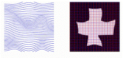
Deformation
Field A
"map" of what stretching, squishing, moving and resizing operations
need to be done to each voxel so that the individual brain you are normalizing can be fitted to
the template brain. This deformation field is generated as an *sn.mat file
by normalization in spm and can be used for vbm.
Here we see a representation of a simple deformation field (left) and then
we see the field applied to an image of a cross to warp it (Image from:
Ashburner, J and Friston, K.J. "Spatial Normalization using Basis
Functions" Chapter 3, Human Brain
Function)
Depth See Image Depth
Design
matrix A
model of your experiment and what you expect the neuronal response to it to
be. In general represented as a matrix (funnily enough), where each row
represents a time point / TR / functional image and each column represents
a different experimental effect. It becomes the model in a multiple
regression, following the vector equation: Y = BX + E. Y is a vector of
length a (equal to nframes from the scanner), usually representing the
signal from a single voxel. B is a vector of b, representing the effect
sizes for each of b experimental conditions. E is an error vector the same
length as Y. X is your design matrix, of size a x b. (From Gablab
Wiki: Glossary)
DICOM (Digital Imaging and Communications in
Medicine) The DICOM image format is commonly used for transfer and storage
of medical images. Visit Chris Rorden's Dicom
page for information about the format and free software to view and
manipulate it. See also Image, ezDicom and MRIcro.
Diffusion Weighted Imaging (DWI) MRI sequences
weighted by the diffusion of water. Diffusion Weighted Imaging (DWI)
measures the molecular mobility of water in tissue. Less attenuation of
signal is expected in regions of less restriction (i.e., less compartmentalization
of the water). Diffusion in biological systems is complex, but directly
related to tissue microstructure, which differs in normal and diseased
tissues. Diffusion weighted MRI can be used to diagnose diseases like
stroke and multiple sclerosis.
Diffusion Tensor Imaging (DTI) refers to diffusion imaging
using Tensors (A mathematical construct for describing
multidimensional vector systems). Diffusion Tensor MRI can be used for
mapping white matter tracts. The acronym ADC refers to the Apparent Diffusion
Coefficien (a quantitative measure of diffusion based on Diffusion Tensor
Imaging), a quantification of diffusion. See also isotropic
and anisotropic. See DPTools. See also the following
interesting links:
A
master's thesis with good descriptions and images of the basics of
diffusion. Particularly helpful are the contents links on the first page:
Image Acquisition and Tensors.
http://www.rsierra.com/DA/thesis.html
A good diffusion introduction in Nature Reviews Neuroscience:
http://www.nature.com/cgi-taf/DynaPage.taf?file=/nrn/journal/v4/n6/full/nrn1119_fs.html
Other
software that may be useful: http://midag.cs.unc.edu,
http://www.poldracklab.org/spm/spm_dti.html
(seems to be for Siemen's images)
http://rsl.stanford.edu/research/software.html
(A variety of potentially useful programs, including one to convert ge
images to dicom).
See
also http://www.dtiatlas.org/
, an atlas of DTI images.
Dilation This
operation gradually increases the area of foreground pixels in an image.
See HIPR's Dilation
Page. Surefit uses dilation to help correct topological defects in
a reconstruction. See also erosion.
Dispersion derivative
The derivative with respect to the dispersion parameter in a gamma
function. In SPM, the dispersion derivative of the canonical HRF looks a
lot like the HRF but can be used as a basis function, to model some
uncertainty in how wide you expect the HRF to be at each voxel.(From Gablab
Wiki: Glossary)
Distal Away from the core or center. Compare to proximal.
DMDX A program
written by Jonathan and Ken Forster. The program runs under MSWindows and
is used to present a series of precisely timed stimuli to a viewer. Check
out the DMDX Updates Page for several useful links: http://www.u.arizona.edu/~jforster/dmdx.htm.
See also Presentation.
If
you use DMDX in your project, you should probably reference the following
paper:
Forster,
K.L., Forster, J.C. (2003). DMDX: a windows display program with
millisecond accuracy. Behav Res Methods Instrum Comput.35(1):116-24.
Dorsal Toward the back. In the case of the
brain/head, used to mean toward the top of the head, same as Superior. Compare to ventral or inferior.
Downloads Download links for this site can be found in several
places. Here are some primary downloads:
DPTools MS
Windows software designed to aid in diffusion
and perfusion analyses. See the
fmri tools Home page. See
also Activ2000.
DTI See Diffusion. See
also http://www.dtiatlas.org/.
DWI See Diffusion.
E
Epoch See Block Design
Erosion This operation erodes or shrinks foreground pixels causing
holes within those areas to become larger. See HIPR's erosion page
for more information. Erosion is used by Surefit
to correct topological errors. See also dilation.
Errors (at the scanner and with the presentation equipment) To
submit reports of errors during scanning, please use the following subject
lines for, respectively, problems (and maybe solutions) on MR1, MR2 and
MR3, and likewise problems and/or their solutions for the presentation
system (including goggles, cables, stereo, hercules etc):
MR1
problem
MR2 problem
MR3 problem
Presentation problem
Euler
Count An
automated count of topological errors based on the algorithms of Leonhard
Euler.
Event-Related Design An experimental design in
which the time a stimulus is presented is not fixed and is often (but not
necessarily) extremely brief. Also sometimes called Single trial design
because each trial is statistically independent of the other trials. To be
statistically independent, different trial types must be intermixed so that
it is not possible to predict the next trial type from the previous one.
(Compare to Block Design, SPM99WorkbookStudy1.doc
See also Afni-MGH Event
Related Analysis, MGH).
Event Related Tutorial: SPM99event.doc
EzDICOM The free ezDICOM
software runs on Windows computers. It is able to display most
types of DICOM image (many other viewers are limited to showing
uncompressed grayscale DICOM images) and can automatically detect and open
Analyze, DICOM, Genesis, Interfile, Magnetom, Somatom and NEMA images. If
MRIcro doesn't open the image, you should try ezdicom. For example, a
single slice from one of our P-files can be opened in ezDicom as
follows:File=>Open Raw: Width 64, Height 64, Slice Frames 1, Offset 0,
Bits per Pixel 16, Convert Raw to Analyze, Dicom or interfile (you choose).
F
F-contrast Simply put, a T-contrast tests a single linear
constraint on your model - something like "The effect size (parameter
weight) for condition A is greater than that for condition B."
T-contrasts can involve more than two parameters, but they can only ever
test a single sort of proposition. So a T-contrast can test "The sum
of parameters A and B is greater than that for parameters C and D,"
but not any sort of AND-ing or OR-ing of propositions.
An
F-contrast, by contrast (ha!), is used to test whether any of
several linear constraints is true. An F-contrast can be thought of as an
OR statement containing several T-contrasts, such that if any of the
T-contrasts that make it up are true, the F-contrast is true. So you could
specify an F-contrast like "parameter A is different than B; parameter
C is different than D; parameter E is different than F," and if any of
those linear contrasts were significant, the F-contrast would be
significant. The utility of the F-contrast is highest when you're just
trying to detect areas with any sort of activation, and you don't
have a clear idea as to the shape of the response. They were designed to be
used with something like a Fourier basis set model, where you want to know
if any combination of your cosine basis functions is significantly
correlated with the brain activation. Testing that set with a T-contrast
wouldn't be correct; it would tell you whether the sum of those basis functions'
parameters was significant, which isn't what you'd want. Testing
individually whether any of those parameters is significant, though, tells
you something.
The
disadvantage of the F-test is that it doesn't tell you anything about which
parameters are driving the effect - that is, which of the linear
constraints might be individually significant. It also doesn't tell you
what the direction of the effect; parameter A might be different than
parameter B, but you don't know which one is greater. This isn't a problem
if you're using a basis set where different parameters don't have much
individual physiological meaning (such as a Fourier set), but oftentimes
F-tests are followed up with t-tests to further isolate which parameters
are driving the effect and what direction the effect is in. (From Gablab
Wiki: Contrasts FAQ )
FFT Fast Fourier Transform A method for deconvolving
(taking apart) any complex waveform into its component sinusoidal
waveforms, which aids greatly in their analysis. Used in acoustic analysis,
fMRI, seismic analysis and lots of other things.
Fiasco An free fMRI analysis package
for unix developed at the Statisitics dept, University of Pittsburg.
It is a series of shell scripts and executables for several unix platforms.
Some associated tools have been developed in Java and fiasco interacts with
R (It is not clear whether it is at all dependent upon R). Fiasco uses its
own proprietary "pgh" file format, described here.
See http://www.stat.cmu.edu/~fiasco/
FICO Afni data
type: Functional Intensity+ Correlation data (Multibucket)
Field
of View see FOV
File system The way in which an
operating system organizes its files and directories
File Transfer A page describing machine
specific file transfer protocols for the CNL
Filter Filters
remove unwanted frequency components from signals. Some filters preserve
low frequencies but remove high frequencies (low pass filters).
Others do the opposite (high pass filters). A band pass
filter passes selected frequencies while cutting out others (e.g. You could
produce a bandpass filter by doing both lowpass and highpass filtering--as
long as you let some frequencies through). Several different filter shapes
are available for smoothing images. In general, the the center voxel is
weighted more heavily in a smoothing operation. However, the shapes of the
filters vary. Blackman filters are relatively leptokurtotic
(narrow), Hanning filters are intermediate and Hamming
filters are the fattest. Gaussian filters are typically lowpass
filters, used for smoothing in spm, Cambridge
Introduction to Smoothing. The width of the Gaussian is sigma (the FWHM).
FIM Afni type: Functional intensity Map.
The file is actually 3d because time data is merged.
Find A powerful unix command with
difficult syntax. It can be used to find files or directories by ownership,
age, name etc. and do almost anything with them (change them, delete them
etc.)
FIR
(or Finite Impulse Response) model A type of design matrix which assumes nothing
about the shape of the hemodynamic response function. With an FIR model,
you don't convolve your design matrix with a canonical HRF or any basis
functions. Instead, you figure out how long an HRF you'd like to estimate -
maybe 10 or 15 TRs following your stimulus. You then have a separate column
in your design matrix for every time point of the HRF for every different
condition. You separately estimate beta weights for every time point, and
then line them up to form the timecourse of your HRF. The advantage is that
you can separately estimate an unbiased HRF at every voxel for every
condition - tremendous flexibility. The disadvantage is that the confidence
in any one of your estimates will drop, because you use so many more
degrees of freedom in estimation. Full FIR models may not be useable for
very complex experiments or certain types of designs. (From Gablab
Wiki: Glossary)
Fitted Response (In SPM)
Simple plot of mean (averaged over session) regressor across PST.
FLAIR Fluid Attenuated Inversion
Recovery
Flat
mapping The process of rendering all or part of the gray matter,
whether it is folded into sulci or gyri, as a flat surface accessible in a
single view. See also Surface Based
Analysis, Mrgray, Surefit/Caret,
Freesurfer and BrainVoyager.
fMRI (Functional Magnetic
Resonance Imaging) offers the cognitive neuroscience community enormous
promise for understanding both normal and pathological brain functioning.
The technique provides measures of regional change in brain activity while
subjects are engaged in various cognitive tasks, combining activation
information from the functioning brain with the exquisite anatomical detail
of high-resolution structural MRI. Unlike other functional brain imaging
techniques, fMRI is a completely noninvasive procedure; subjects are not
exposed to chemical agents, radioactive materials, or X-rays. The benign
nature of the procedure allows the implementation of longitudinal research
designs since the same subject can be tested and re-tested over multiple
sessions; a distinct advantage in longitudinal research aimed at following
normal and abnormal development of cognitive functioning across the life
span. Other functional MRI techniques, such as perfusion arterial spinal
labeling MRI, diffusion-weighted MRI, and MR spectroscopy, are currently
being developed that will greatly enhance our ability to measure brain
functioning, especially in the presence of neuropathology. MRI techniques
such as these will undoubtedly result in important contributions to
clinical sciences as well, in the areas of diagnosis, assessment of risk,
treatment monitoring, and drug development. For these reasons, fMRI is
likely to become the dominant tool for examining brain function in humans.
See also functionals, and k-space.
Font Sometimes
a specialized font is used for experiments. To use a font, you should put
it in the font area on windows (go to your C drive and search for
"font"). On Windows 2000 it is C:\WINNT\Fonts. You can download
the backward font here.
It is called ARIALBK.TTF. (TTF=True Type Font).
Format Image file formats used by programs that
process medical images come in a somewhat depressing variety of flavors.
Essentially, each program has its own format AND there are several
competing standards. To begin with, MR scanners produce images in a variety
of proprietary formats (see Genesis,
the GE format). The following formats used by various medical image
processing programs are also of particular interest:
- bfloat/hdr
(a format used for functional data in the MGH-fsfast
software)
- BRIK/HEAD
(used by Afni),
- bshort/hdr
(a format sometimes used for structural/anatomical files by the
MGH-fsfast software),
- COR
(the native freesurfer/mgh
format for structural/anatomical file),
- dcm
(dicom format-an attempt at standardization of a medical image
format),
- img/hdr
(Analyze format, sometimes called the AVW format, used by AIR, FSL,
Medx, Spamalize, SPM, and VIDA with slight modifications...they
"should" all be compatible),
- mnc
(the minc format-another attept
at a standard, supported by afni and used by surefit)
See Image and Conversion and nifti.
FOV Field of
View, the size, in mm of the space (i.e, real distances) represented by the
image (voxel/pixel size x number of voxels/pixels).
For example, an image with 10 isotropic pixels, each 5 mm square, would
have an FOV of 10x5 or 50 mm. Field of view can, of course, be different in
each direction.
Freesurfer A
free image manipulation program from the Massachucets General Hospital.
For the most up-to-date and well-tested copy of freesurfer and the new mgh
tools, use zoe, and see the mgh page for
further information on setting up your environement and data to use the
programs. Freesurfer uses the COR
file format. See sample images at Marty
Sereno's website, and MGH. http://www.nmr.mgh.harvard.edu/freesurfer
. See also mgh, tkmedit, and tksurfer, and nifti.
from3d A file conversion program
that comes with the afni package.
FS-FAST See MGH
FSE Fast Spin Echo, a pulse sequence
characterized by a series of rapidly applied 180° rephasing pulses and
multiple echoes, changing the phase encoding gradient for each echo. http://fonar.com/glossary.htm
FSL The FMRIB
Software Library (FSL) is a collection of functional and structural brain
image analysis tools, written mainly by members of the Oxford Image
Analysis group. These are stand alone binaries available for several
platforms. Type >fsl on Merlin, Holly or Charlie, where the program
is installed. fsl uses the AVW (Analyze img/hdr) format. See also nifti. Visit http://www.fmrib.ox.ac.uk/fsl/index.html
for download, online documentation of the tools, etc.
Note: Your .cshrc must have the
following 2 items for fsl to run for you on merlin, holly or charlie:
/usr/local/bin/fsl in your .cshrc path
and the following environment variable line:
setenv FSLDIR /usr/local/bin/fsl;
FTP (File Transfer Protocol). See
also specific protocols for transfer
between campus and UMC or from the console to our UMC workstations. (see
also transfer, special
techniques for transferring images back to the console; See Trouble
with permissions)
Functional
files P-files (fMRI, "functional magnetic resonanace
images") record the spatial and temporal coordinates of changes in the
brain's blood oxygenation levels in k-space.
A P-file is usually a series of thousands of images (shorts with 16 bit image depth, in our case). Each image
is a single slice through the
brain. Because each slice has thickness, the images are made up of voxels rather than pixels. Our voxels are typically about
3.5 mm x 3.5 mm x 5mm. Our image slices are usually in axial. Images have a radiological orientation. This
relatively low resolution
limits our ability to pinpoint the locations of activations. Each slice is
64x64 voxels and a set of approximately 17-19 slices will represent a brian
volume (nas, number of anatomical slices). Depending on the length of the
experiment, we may record from ~80 to ~250 brain volumes (nfs, number of
functional slices; aka, ntr). During
preprocessing, Afni treats these
P-files as low resolution 4D anatomical files (although they contain
functional information). As part of analysis, the files become 3d fim and fico
files. See also Anatomical file,
and ezDICOM. See rdgehdr (Read GE Header).
FWHM (Full
Width half Maximum) A criterion for defining the width of a curve. The
width of the curve is its width at half its maximum height. See also Half maximum.
G
Gablab Resources Check out Jeff Coopers Wiki pages at http://gablab.stanford.edu/docs/.
Log in as fmri, password fmri. The wiki is a collaborative effort to create
a useful repository of shared information for Neuroimagers.
Garage
Reservations
See Reservations.
Gaussian
Filter see filter
GE General Electric
Medical Systems Home page
General
Linear Model
The general linear model is a statistical tool for quantifying the
relationship between several independent and several dependent variables.
It's a sort of extension of multiple regression, which is itself an
extension of simple linear regression. The model assumes that the effects
of different independent variables on a dependent variable can be modeled
as linear, which sum in a standard linear-type fashion. THe standard GLM
equation is Y = BX + E, where Y is signal, X is your design matrix, B is a
vector of beta weights, and E is error unaccounted for by the model. Most
neuroimaging software packages use the GLM as their basic model for fMRI
data, and it has been a very effective tool at testing many effects. Other
forms of discovering experimental effects exist, notably non-model-based
methods like principal components analysis. (From Gablab
Wiki: Glossary) generic2bshort One of a suite of imaging
tools from UCLA. This one is used to convert a series of *.MR files into a
bshort file. The tools are installed on Charlie, but not yet well tested. See UCLA Brain
Imaging Center.
Genesis A file
format (*.MR) produced by the General Electric MR Signa 5X. This is the
native format saved by the Signa MR scanner. The data are stored as short
integers (no fractional parts), with a single file for each image (a single
location or time point). The first 7904 bytes of the file contain header
information. (UCLA
Brain Mapping Center Image Format Page)
genesis2analyze One of a suite of imaging
tools from UCLA. This tool converts *.MR files to img/hdr files. The tools
are installed on Charlie, but not yet well tested. See
UCLA Brain Imaging Center.
Getting Started What you need to know to
create or help run an fMRI experiment.
Ghostview and Ghostscript Programs for viewing and printing of postscript files. Ghostscript
contains the necessary fonts etc to support Ghostview. Ghostview is the
actual reader. The programs are available for unix, macintosh and PC
platforms. http://www.cs.wisc.edu/~ghost/
Global
effects
Any change in your fMRI signal that affects the whole brain (or whole
volume) at once. Sources of these effects can be external (scanner drift,
etc.) or physiological (motion, respiration, etc.). They are generally
taken to be non-neuronal in nature, and so generally you'd like to remove
any global effects from your signal, since it's extremely unlike to be caused
by any actual neuronal firing. (From Gablab
Wiki: Glossary)
Global
scaling
An analysis step in which the voxel values in every image are divided by
the global mean intensity of that image. This effectively makes the global
mean identical for every image in the analysis. In other words, it
effectively removes any differences in mean global intensity between
images. This is different than grand mean scaling! Global scaling (also called
proportional scaling) was introduced in PET, where the signal could vary
significantly image-to-image based on the total amount of cerebral blood
flow, but it doesn't make very much sense to do generally in fMRI. The
reason is because if your activations are large, the timecourse of your
global means may correlate with your task - if you have a lot of voxels in
the brain going up and down with your task, your global mean may well be
going up and down with your task as well. So if you divide that variation
out by scaling, you will lose those activations and possibly introduce
weird negative activations! (see the Gablab Wiki PhysiologyFaq for some),
considering that moment-to-moment global variations are very small in fMRI
compared to PET. They can be quite large session-to-session, though, so
grand mean scaling is generally a good idea (see below). (From Gablab
Wiki: Glossary)
GNU
Licensing Afni is now distributed under the GNU
Open Source Free software license. Essentially, this license says that you
can freely distribute a product and incorporate it into anything you build
BUT, as soon as you incorporate it into something you build, you can no
longer sell your product, but must also distribute it freely. http://www.gnu.org/copyleft/
Goggles The
new goggles from Resonance
Technologies require different setup than the old goggles. See our
first draft of instructions: new3tgoggles2.doc
. See also the Machine page for updates
on the goggles, and Resonance
Technology (below) for contact information. See information on Hercules, the mobile presentation
machine and Pharaoh, the 3T presentation
machine.
One
presentation system is permanantly installed on the 3T scanner. It includes
a goggle/audio presentation system from Resonanace Technologies. The
goggles are VisuaStim digital goggles with 3d stereoscopic capability, and
an eye tracking system. The audio system is stereo with two way
communication. The presentation computer is a Dell Dimension 8300 running
Windows XP Prowith a 3.0 Ghz processor, 2 GB of main ram, a large SATA hard
drive, an NVIDIA GeForce FX 5200 video card with 128 mb of ram onboard and
3d-stereo capability. The system uses custom hardware and a Measurement
Computing PCI-DIO card to gather responses from two mice used by subjects
in the scanner. In addition, this custom hardware setup allows researchers
to automatically start the scanner sequence with a TTL pulse.
Grand
mean scaling
An analysis step in which the voxel values in every image are divided by
the average global mean intensity of the whole session. This effectively
removes any mean global differences in intensity between sessions. This is
different than global scaling! This step makes a good deal of sense in
fMRI, because differences between sessions can be substantial. By
performing it at the first (within-subject) level, as well, it means you
don't have to do it at the second (between-subject) level, since the
between-subject differences are already removed as well. This step is
performed by default by all the major analysis software packages.(From Gablab
Wiki: Glossary)
Grant Proposal
Writing See Grant
Proposal Writing
Grecons This is
a c program from Gary Glover at Stanford (we actually have several
different versions for different sacanner output) that constructs images
from our raw GE spiral fmri files (P-files). We have versions of grecons5x
for the sgi and sun solaris. See spirec.
See also Scanner Updates for information
about the post September 2002 version of grecons AND information on the
November, 2003 spiral code upgrade on the 3T. See the Grecons Table to determine
which version of grecons you need to use. See prep
for information of shell scripts that will make your life easier.
grecon2bshort An image conversion script
that comes with the mgh package. Very similar to afnireg2bshort.
grhyp reconstructs spiral diffusion
images acquired in 43 directions. It is installed on buddy, holly and
merlin. The command to run it is grhyp [rawfile] [prefixforoutputfile] ,
e.g.,
>grhyp P12345 study
Type
>grhyp
for help
Group
Analysis For
details of running group analysis in spm, see SPM99WorkbookStudy1.doc
Gui a graphical user interface on
a computer, where you use the mouse to move around and click on things.
Gzip (also gunzip to
decompress a gzipped file). Because gzip only acts on files, you may also
find it useful to look at a find
command that will recursively gzip files in multiple subdirectories.
Software like Iceows from http://www.iceows.com/HomePageUS.html
will work on Windows systems to ungzip and untar files that might otherwise
be difficult to handle.
H
Handle A topological error, sometimes called a
"crossover", in which a surface reconstruction is folded over
onto itself. A "bridge" of gray matter across a sulcus is called
an exohandle. A hole through the white matter between two sulci is an
endohandle. Concern over topological errors arises in gray matter reconstruction routines
such as those used in Surefit and
Mrgray. See also cavity and topology.
Half
Maximum (half max criteria) A notion frequently used in image
processing to define an edge. The edge is said to occur when you are 1/2
way between the brightest and darkest points along a line that crosses that
edge. (Keep in mind that you see an edge in a grayscale image because there
is something bright next to something darker). See also FWHM
Hamming
Filter see filter
Hanning
Filter see filter
HDR Hemodynamic
Response, the BOLD (blood oxygen level dependent) response caused by
activity in the brain and measured by fMRI. (See also hrf)
Header Descriptors that accompany an
image and define its format. See Image
and Offset and rdgehdr (Read GE Header).
Help How to find help for unix
Hercules
See also setup
High Pass filters see filter
History A listing of commands that you
recently typed into a unix shell.
Horizontal (same as
"transverse" and "axial")
HRF The
hemodynamic response function. A mathematical function representing the hemodynamic response. May be used
synonymously with hdr (they are very close).
Human
Subjects To
work with people, you must pass a test demonstrating that you know the
rules of interacting with and experimenting on people. The main Human Subjects Protection Site
for the U of A. Link to online
test and other training materials.
I
IDL The Interactive Data Language, is software for
data analysis, visualization, and cross-platform application development.
Type >idl at the command prompt on Merlin where it is installed;
or to start IDL with the GUI, type >idlde at the prompt on
Merlin. See also and look online for tutorials (there is a lot out there).
Useful Links: IDL Home, Program
Library, Spamalize
IATR The goal for this site is to
provide a centrally available listing of all image analysis tools that are
available to the neuroscience community in order to facilitate the development,
identification, and sharing of tools that are of use to the general
community. It is hoped that this helps the "tool developers" to
get their tools to a larger user community and to reduce redundancy (or at
least utilize tool redundancy to facilitate optimal tool design) in tool
development. This also helps "tool users" in identification of
the existing tools for specific problems as they arise.
IID (Independent and Identically
Distributed) The assumption of a statistical model that errors are
indpendent and identically distributed.
Image The
images we get from the scanner are all bitmap data. Such images consist of
a set of numbers that specify the colors of each individual pixel (or
voxel) in the image. A simple bitmap file consistes of a header and the raw
bitmap data. The header may be stored internally to the image (as is
the case for our raw MR files) or externally, as is the case for the
Analyze-like format used by SPM (For each image there is a *.img and a
*.hdr file). The header may contain information about the palette of colors
used in the image (LUT), a file
identifier (a code that tells the computer what kind of image it is, e.g.,
*.bmp, *.jpg etc), the number of lines per image, the number of pixels per
line, the number of bits per pixel, compression type, x,y, and z origins
etc. Image conversion usually involves changing the header information to
match the expected order and position of elements in the desired image
type. For details, see Anatomical
Files, Functional Files,
Offset, Pixel, Resolution and Voxel. Learn more about medical image
file formats: Analyze, DICOM, MINC,
NEMA, nifti,
VTK,
Afni Brik, MGH bshort, MGH bfloat. Look at the Medical Image
format FAQ. Learn more about Image
Processing and Image Properties.
Learn about conversion between formats.
Look at information about the mgh program mri_convert.
Image
Capture See Screenshots
Imaging_analysis
listserv The listservs will be replaced by the webboard by the end of January
2005.
ImageJ A
cross platform Java based version of NIH Image. Type >ImageJ on
Merlin to access it. http://rsb.info.nih.gov/ij/
Image
Depth The number of colors or levels of grey scale that can be assigned
to a single pixel or voxel. A binary (one bit) image can have two colors
(usually black and white). You will also commonly encounter 8 bit images
(2^8 or 256 colors or levels of grey) and 16 bit images (i.e., 2^16 or
65,536 colors or levels of gray). A 16 bit image takes twice as much room
as an 8 bit image to store in a computer. A binary image takes very little
room to store. See also Anatomical
Files
imcalc A
function in spm
which allows you to perform basic algebraic operations on images. It is
analogous to 3dcalc in afni. For
example, imcalc will allow you to perform Matlab dot multiplication on two
images with a command like this to imcalc:
i1
.* i2
(where
i1 is the first image you select in the spm interface and i2 is the second
image).
imconvert one of a suite of tools available from the UCLA Brain Imaging Center.
Inferior
Toward the underside of the brain or head. Same as ventral (which means
toward the stomach as opposed to the back)
inorm An MGH program that
calculates the mean intensity data for the entire functional volume. The
global mean of the fMRI signal inside the tissue is calculated by
segmenting tissue from air. The number is later used to rescale the data so
that when intersubject averaging is done, all subjects have the same global
mean. inorm.ps
Interpolation The process of computing new
intermediate data values between existing data values. (From Gitta Domik's Tutorial
on Visualization). Interpolation may be used to smooth out jagged
corners and edges on activation voxels, for details on doing this in Afni,
click here.
Isotropic
Exhibiting equal physical properties or actions (OED).
A
characteristic of a pixel or voxel that is square or cubic
respectively. Compare to non-isotropic.
See the Caret (for Pfile/Functional files)
and Surefit (for 2D and 3D anatomical
files) pages for instructions on converting non-isotropic voxels to
isotropic voxels.
In diffusion weighted imaging,
isotropic movement of water molecules is movement that is unimpeded, or
equal in all directions. Compare to anisotropic.
isxavg an mgh program isxavg.ps
itk-snap A free image segmentation and
roi creation tool.
J
Jitter
"The practice of varying the timing of your TR relative to your
stimulus presentation. It's also often connected to, or even identified as,
the practice of varying your inter-trial interval. The idea in both of
these practices is the same. If your TR is 2 seconds, and your stimulus is
always presented exactly at the beginning of a TR and always 10 seconds
long, then you'll sample the same point in your subject's BOLD response
many times - but you might miss points in between those sampling
points" (Gablab Wiki). By
varying the relationship between the experimental condition and TR we can increase the success of
deconvolving the FFT (signals) for
each experimental condition from the other conditions. This becomes
especially important when trials are very short (e.g., a few seconds). Optseq can be used to calculate how
to add appropriate jitter to an experiment.
K
K-space Raw or Time Domain data used for raw functional files. A data map
based on signal amplitude versus position rather than direct position
within the subject. kspace.ppt
(If you have powerpoint, you can watch a virtual reconstruction of k-space.
Simply click the link, then each mouse click on the image will update the
reconstruction). See the animated tutorials on kspace here MRI Q&A
for Physicists: Kspace
Khoros
and Cantata A high level programming interface in which one can
attach widgets to one another in paths, modify their characteristics, and
run signals (like images) through the entire path to filter or modify them.
http://khoros.com/
L
Lab
Manual Online (CNL Lab Manual)
Left
Handed Coordinate System Left is right and right is left in viewed images. See
radiological orientation. Left
Lesion Test Image The Left Lesion
Test Data (This data has a big hole in the left front, so you can
test your understanding of what is happening to right and left given a
particular program or manipulation. There is a single functional image, and
"2D" and "3D" structural images in spm format).
Less A viewer, much easier to use and more flexible
than more. Less is the default viewer for man pages on buddy, holly, merlin
and charlie.
Linear
Transformation See Affine
Transformation
Link A link is
like a shortcut under windows. You can make what is called a soft link by
doing something like the following:
ln
-s /data/harvey/run1/joe fred
This
will create a link called fred in your current directory (the one you are
in when you type the command). The link will point to /data/harvey/run1/joe. If you
cd to fred, you will end up in /data/harvey/run1/joe.
If you rm fred at some later time, fred will disappear, but /data/harvey/run1/joe will
remain.
Library
Listserv The listserv will be replaced by the webboard by the
end of January 2005. See webboard
in this glossary for more information.
Little
Endian Describes a computer architecture in which, the least
significant byte of a multibyte numeric representation is stored in the
lowest-memory address, which is the address of the data. PCs use this
format, whether thay are running Linux or Windows. See also Big Endian and Byte swapping.
Low
pass filter
see filter
LUT Lookup
table. Pairs of numerical values that allow a program to match a meaningful
value to one which specifies a color on the output device.
M
Machines
Magnet See Scanner
Marching
Cubes A method of visualizing 3-D data structures by looking for
level surfaces in a 3D-space comprised of a lattice of points. In contrast
to volume rendering, where one can see the entire structure, marching cubes
only allows a single surface to be rendered (From Gitta Domik's Tutorial
on Visualization). Marching Cube Algorithms are used in Mrgray and Surefit to construct the gray matter
surface.
Marsbar An SPM ROI Toolbox developed by Matthew Brett. See also SPM and MarsbarNotes.doc
If you use Marsbar in a paper, use the following reference:Matthew Brett,
Jean-Luc Anton, Romain Valabregue, Jean-Baptiste Poline. Region of interest
analysis using an SPM toolbox [abstract] Presented at the 8th International
Conferance on Functional Mapping of the Human Brain, June 2-6, 2002, Sendai, Japam. Available
on CD-ROM in NeuroImage, Vol 16, No 2.
Mask In general,
a mask is a file that filters out values from another file. The simplest
way to do this is to draw shapes on an image and use the shapes to define
what values are seen in some other image. Typically, the mask image has 1's
inside the shapes and 0's outside. When you multiply an image by this 1,0
mask, only the values multiplied by 1 remain. The others all turn to 0's.
Afni and SPM have associated mask drawing tools. Massachusetts General Hospital
See also MGH and Freesurfer.
Matlab
(Matrix Laboratory). A mathematical programming
language and environment, optimized for matrix operations. Matrix
operations are crucial to all kinds of signal processing for both sound and
images. Matlab programs are *.m files, which are plain text scripts. Matlab
matrix data is stored in *.mat files. type >matlab to start the
gui interface, or >matlab -nojvm to start the command line
version of matlab on Merlin, buddy or Holly. http://www.mathworks.com/. See
also Matrix, Cell Array, and Vector.
Matrix MEDx A unix based commercial
medical image processing application from Sensor
Systems
mc-sess An
mgh script that runs the afni motion correction.
Merlin
MGH Massachusetts General
Hospital produces a suite of image processing programs, primarily
for event-related analysis. The new suite (see #2 below) includes afni,
fsl, freesurfer and the fs-fast tools. See also freesurfer, tkmedit, and tksurfer. IMPORTANT : We use
two distinct versions of the MGH utilities at the CNL:
1) The older version which does NOT include Afni,
FSL, Minc tools or Freesurfer. See pages on afni
preprocessing and afnievent analysis
for use of the old mgh program.
2) The new (but not necessarily better) MGH
program only runs on linux. It DOES include afni, freesurfer, minc tools
and fsl. It is not something we have been able to use very successfully
yet, though we have tried and have some documentation on installing and
using it on the MGH page.
Citations for those using the mgh event related
processing stream:
Glover, G. H. & Lee, A. T. (1995). Motion
artifacts in fMRI: comparison of
2DFT with PR and spiral scan methods. Magnetic
Resonance in Medicine, 33,
624-635.
Dale, A. M. (1999). Optimal experimental design for
event-related fMRI.
Human Brain Mapping, 8, 109-114.
Dale, Greve & Burdock ???
MINC (Medical Image NetCDF) is built on the general data format NetCDF
(from the UCAR - University Corporation for Atmospheric Research). The MINC
format was developed by the Montreal
Neurological Institute. See also Surefit
and Afni.
mkcontrast is an mgh
program mkcontrast.ps
MNI A template brain based on the average of a large set of brains
(see also talairach). The
template was developed by the Montreal
Neurological Institute. SPM uses the MNI brain. See also BIC. For some interesting spm99 add on
tools for viewing MNI and Talairach information, visit http://www.ihb.spb.ru/~pet_lab/
mni2tal A freely available matlab
script that converts mni coordinates to talairach coordinates:
Sample Input:
>>mni2tal([40 -16 -30; -45 -73 -12])
Output will appear on the screen and in a file called
tal_out.txt
mni2talb was created to handle MGH
Label files as input, hence it expects 5 values in each row rather than 3.
It then strips off the first and fifth columns.
Both are available here.
http://www.mrc-cbu.cam.ac.uk/Imaging/Common/mnispace.shtml
A discussion of the differences between talairach space and mni space, and
links to matlab files to translate from one to the other.
Montreal
Neurological Institute See also MINC
and MNI.
Morphometry
"Shape measurement", a term used to describe measurement of brain
structures (like the hippocampus, amygdala etc.), generally for group
comparisons.
Move move files
move directories
Movie It is possible to make a
simple movie (rotating brains etc.). For Afni,
see Volume Rendering.
MR files (see Anatomical files)
MR1, MR2 and MR3 These are
the 3 MRI scanners. Information about them can be found in two places: The Scanner Updates page (descriptions, safety,
scheduling, analysis implications of using each machine, links to the
MRITech Manual) and the Machine page
(current status). See also transfer
(special techniques for transferring images back to the console). To
understand how number of slices
interacts with number of repetitions
and TR, see Spiralio Sequence Limitations.
MR2bshort (and
MR2bshort2) CNL image conversion utilities that are based on a script that
belongs to the mgh package.
mri_convert An MGH
binary that does considerable image conversion. Can be found on
Charlie...largely untested.
usage: mri_convert [options] <in volume> <out
volume>
Mrisun
MrGray MrGray and MrFlatmesh work together to produce
flattened represenatations of gray matter. MrGray is a standalone
executable that runs on MSWindows. MrGray comes with a program to
convertAnalyze/SPM headers to Mrgray's *.dat image files. MrFlatMesh is a
set of Matlab routines that will run on any platform running Matlab. http://white.stanford.edu/~brian/mri/segmentUnfold.htm
MRI Magnetic
Resonance Imaging See http://fonar.com/glossary.htm
for a glossary of related terms. See the Links page
for educational references. See also http://www.mr-tip.com
MRICro A free Windows Image Viewing and manipulation program:
http://www.mrc-cbu.cam.ac.uk/personal/chris.rorden/mricro.htm
See also Dicom and NEMA and MRIcron.
Multibucket image An image that has more than one value per pixel/voxel. e.g., In afni, an intensity fMRI image (fim)
that has been correlated to behavioral changes (fico). Both the intensity
and the correlation values are stored in each voxel. See buckets.ps.
See advice on splitting a
multibucket image.
N
nas Number of anatomical slices
in each brain volume of a P-file/Functional
image.
NEMA (National
Electrical Manufacturers Association) An organization associated with
medical image standards,the ACR/NEMA
1.0 and 2.0 Standard is their earlier standard. The ACR/NEMA
Dicom 3.0 is the more recent standard and is more accepted by the
community. See also DICOM, MRIcro and Image.
Neurological Orientation Left is Left and right is right in a displayed image.
This is also called the Right handed Coordinate system. (Compare to Radiological Orientation).
nfs Number of functional slices
(number of brain volumes) in a P-file/Functional
image. See "number of
repetitions".
NIfTI Neuroimaging Informatics Technology
Initiative An up and coming image format (as of 5/04/2004) being
developed by a consortium including those who develop afni, spm
and fsl, freesurfer (and others). The idea
is to support a shared image format that will allow researchers to move
data more easily between image processing programs and to have an optimal
format for backup.
NIH Image A
generalized free Image Processing program that was designed to run on the
Macintosh. It has spawned offshoots: Scion
Image, which runs under MS Windows; and Image J, a true Java based
application that runs on any platform. http://rsb.info.nih.gov/nih-image/index.html.
Published research assisted by NIH Image should use a statement similar to
the following in the materials and methods section "... analysis
performed on a Macintosh computer using the public domain NIH Image program
(developed at the U.S. National Institutes of Health and available on the
Internet at http://rsb.info.nih.gov/nih-image/)".
Non-isotropic A
characteristic of a pixel or voxel that has x,y,(z) dimensions that
are not pqerfectly equivalent. Compare to isotropic.
Nonlinear
Transformation See Affine
Transformation
Non-sphericity See sphericity
Normalization In the
MGH program (inorm), that we use with Afni,
"normalization" refers to signal intensity normalization. That
is, artifactual variation in the strength of the signal through time (and space?)
is effectively removed or compensated for by an averaging process. When the
term "normalisation" is used in relation to SPM, however, it
refers to the process of warping the shape of the brain to a standardized
template, essentially "talairaching" (see also Talairach and MNI)
ntr See Number
of Repetitions
Number of Repetitions (a.k.a
ntr, nfs "number of functional slices"), number of time frames
[on the console itself]) The number of times the brain gets scanned in an
fMRI experiment (the number of tr's).
Typically, at the CNL, the brain is scanned every second or two for 2 to 15
minutes (depending on the length of the experiment). We thus end up with
the number of scans varying from about 80 to about 450. If you try to scan
the brain more than once per second, you have to sacrifice spatial
resolution. If you scan for a long time (more than 15 minutes), the
resultant image files (P-files) are huge. P-files typically run between
about 20 and 50 mb. Experimenters often collect several separate P-files
from a subject in the magnet, for example, one file before some
manipulation and another file afterwards. See functional file. To understand
how number of slices interacts with
number of repetitions and TR, see Spiralio Sequence Limitations.
O
OCF The
scanner schedule. See Scanning
schedule for more information.
od (octal dump) a unix command
that allows you to see every character in a file -including annoying hidden
ones like nl (new line) and sp (space). This is valuable because the hidden
character can sometimes destroy the functionality of otherwise good shell
scripts. See detailed example here.
Offset If you
want to read the image as a "raw" image in ScionImage, ImageJ
etc., then you need to know the offset. The offset (or "skip") in
bytes is the number of bytes to skip in the file because they are header
bytes (i.e., they contain information about the kind of file in the image,
rather than the actual image data). To calculate the offset, take the size
of the file and subtract the size of the image. The remainder must be
header information. Our files are 138976 bytes. Image size for our files
256x256x2 or 131072 (xdim x ydim x imagedepth). 138976-131072=7904. Offset
is 7904 bytes. See also Anatomical
files.
Operating System
The software that controls the computer hardware (Linux, Irix, Windows 2000
etc.)
Optimal Presentation Order see optseq
Optseq optseq is a program included in the old mgh tools (written
by Doug Greve), and thus is installed on buddy, holly and merlin (simply
type optseq to access
it). It can be used to generate an optimal sequence (hence the name) for
conditions in an event related study. See further explanation here. SPM99 appears to have similar
capability as part of Model Specification (described in section 3.2.1 of
the May 2001 SPM Manual). For detailed advice on using optseq, consult
Siobhan Hoscheidt's excellent optseq.doc. optseq2 is
installed on zoe and charlie. To access optseq2, type:
>mgh
>optseq2
optseq2 uses double dashes (e.g., --ntp) before option
flags; whereas optseq uses only a single dash (e.g., -ntp). optseq2 also
uses lower case tr as a flag, whereas optseq
uses uppercase TR). [Thanks to Chun yu Lin for
noting these differences]. Doug Greve's optseq webpage.
See also jitter.
Orientation (For
Subjects)
outlier One of a suite of imaging
tools from UCLA. This tool provides a way to evaluate outliers. The tools
are installed on Charlie, but not yet well tested. See
UCLA Brain Imaging Center.
Ownership
Overlay An overlay is an image (for
fMRI, usually of activations) that is viewed on top of a high resolution
anatomical image (usually called the underlay).
P-file A file containing a series of raw fMRI
images. See functional file
and ezDICOM. See rdgehdr (Read GE Header).
Parametric vs Non-parametric statistics In
general, parametric procedures have more stringent assumptions about the
distribution and character of the variables than nonparametric statistics.
Non-Parametric tests are often used in place of their parametric
counterparts when certain assumptions about the underlying population are
questionable. For example, when comparing two independent samples, the
Wilcoxon Mann-Whitney test does not assume that the difference between the
samples is normally distributed whereas its parametric counterpart, the two
sample t-test does. Non-Parametric tests may be, and often are, more
powerful in detecting population differences when certain assumptions are
not satisfied. All tests involving ranked data, i.e. data that can be put
in order, are non-parametric.
Parfile (parameter
file--used by mgh tools) A text file necessary for event-related analysis.
The file provides information about the stimulus schedule for each run,
that is, details about the times at which each stimulus was presented for a
particular subject (in an event-related paradigm, the timing is dependent
on subject responses). Our parfiles are generally based on an output file
generated by dmdx and then manipulated in a spreadsheet to have the correct
format for the mgh programs. The parfiles are created by the FS-FAST
command optseq.
Paste Unix command for
concatenating text files vertically. See cut
and paste. See also cat.
Path
Perfusion weighted
MRI An imaging technique used for measuring blood flow through the brain,
specifically, the volume of blood supplying a unit mass of tissue per unit
time. Perfusion images can be obtained using a bolus of contrast agent
injected into the patient OR arterial blood can be tagged by magnetic
inversion. Images of the tagged blood flowing into the brain are alternated
with untagged images, and tagged and non-tagged images are subtracted to
remove the signal from brain tissue, leaving the blood signal. See also Introduction
to Perfusion
Peristimulus time (PST) In SPM, the peristimulus time for a given
event is simply defined as the time that has passed since the last
presentation of that particular event type.
Permissions
Phonelist The CNL
phonelist requires a username and password to access. It is in Excel
format.
phx_tospm
A Matlab program (actually, several different versions of the program) that
take greconed, renamed files from our GE scanners and make proper spm img
and hdr files. Unfortunately, the program is prone to being a little cranky
if you don't get everything just right. Get versions of phx_tospm here.
Pilot
Projects Link to the standard CNL form
you need to fill out to apply for pilot project funding. See the Getting Started page too.
Pitch
Pixel A
"picture element", the smallest nondivisible component of a 2D
image. It will have an x and y dimension and a color. (See also bitmap, voxel
and image depth).
Pharaoh 3T
presentation machine.
Planes (of section) The angle of slices. There are 3 standard
angles and as many oblique angles as you can imagine.
PLS (Partial Least Squares
Analysis) The PLS analysis software is free, runs under Matlab and requires
at least the Matlab statistics toolbox (and possibly image processing and
signal processing). It is used to identify task dependent changes in
activity, changes in relations between brain and behavior and functional
connectivity between brain regions. PLS is available for download (after a
brief registration process) from the Rotman Research Institute http://www.rotman-baycrest.on.ca:8080/index.html.
PLS can be used for analysis of PET, ERP or fMRI data. It presumes that the
data have been preprocessed in spm99 or spm2. It assumes the MNI coordinate
system and prefers whole brain scans.
Posterior Toward
the back of the head. Compare to Anterior.
Postscript A page
description language developed by Adobe to allow entire pages to be sent
describing graphics and text to a printer. The flexibility allows more than
just normal ASCII characters to be sent and is the defacto standard in high
end printing. Postscript documents may have the extension *.pdf (Adobe
Acrobat files) or *.ps. You can read Postscript files using xpsview on the
sgis or Ghostview (if it is
installed) on most Unix or PC machines.
Prep Scripts
I have created several unix shell scripts (in
/usr/local/bin) to facilitate data preparation and afni preprocessing. All
are available here
in the tools area on Merlin. They are also available on buddy, holly,
merlin, charlie, pdw_cortex and tommy. Simply type the script name
(assuming it is in the path) and it will tell you how to use it.
Alternatively, you can look at the script in a text editor.
"How-to" info is at the top and various comments appear
throughout to help you understand what the script is doing. Because these
scripts call other scripts and programs, those programs must also be
available. To learn more about versions of grecons, see the grecons table.
- There
are 8 different scripts (prep, prep_v2, prepb, prepb_v2, prepio,
prepio_v2, prepbio, prepbio_v2). prep is the most basic script.
- Versions
with "b" appended are for big data sets (more than 9999
images; i.e., ntr x nas > 9999).
- Versions
with "io" appended are more complex and can only be run on
linux (because we don't have program binaries for the sgis). They are
for the spiral in-out sequence.
- Versions
with "_v2" appended run version 2 of grecons (grecons_v2) ,
and where appropriate (on linux, for spiral in-out sequences, version
2 of expandonefile (expandonefile_v2)).
|
prep runs (and thus is dependent upon the
presence of) grecons and spiral_rename. It also does some cleaning up
(removes B0 files and puts your original P-file in a subdirectory called
Orig).
example:
>prep P12345.7
|
|
prep_v2 is the same as
prep, but runs grecons_v2
|
|
prepb is the same as prep, except it is for big
data sets (i.e., over 9999 images, nfs*nas > 9999). prepb calls
grecons and spiral_renameb.
example:
>prepb P12345.7
|
|
prepb_v2 is the same as prep, but runs version 2 of
grecons_v2.
|
|
prepio depends on grecons, sprlioadd,
expandonefile, and spiral_rename. It assumes 64*64 data, but you could
alter the script if you needed something else (change the 64 in the
expandonefile command to whatever is appropriate). Because sprlioadd and
expandonefile only run on linux and sun, prepio is useless on the sgis.
Use this only if you run the spiralio (spiral-in-out) sequence rather
than the regular spiral sequence. It assumes byte order for the sgi's
Usage: prepio <Pfile> <nfs> <nas> <output
name>
Example: prepio P12345.7 80 17 tap
|
|
prepio_v2 is the same as
prepio, but runs grecons_v2 and expandonefile_v2
|
|
prepbio is like prepio except it runs
spiral_renameb.
Usage: prepbio <Pfile> <nfs> <nas> <output
name>
Example: prepbio P12345.7 80 17 tap
|
|
prepbio_v2 is the same as prepbio, but runs
grecons_v2 and expandonefile_v2
|
|
super
runs prepio_v2 followed by to3d to make a BRIK and HEAD and then
3dAFNItoANALYZE to create correctly oriented img and hdr files assumes
images in axial orientation. Available on linux
|
|
superb
Same as super, but for 10,000 or more files. Available on linux.
|
|
super12
runs grecons12 code (post Sept 15, 2005) but is otherwise the same as
super. Available on linux. Fine for both the quad coil and 8 channel
coil.
|
|
super12_8ch
runs grecons12 code (post Sept 15, 2005) like super12. Uses -G 0.5 to set
the gain for the 8 channel coil to avoid wrap around (causing some washed
out images) Available on linux. Unless you encounter gain related
problems, you don't need this....just use super12.
|
|
superb12
runs grecons12 code but is otherwise the same as superb . Fine for both
the quad coil and 8 channel coil.
|
|
superb12_8ch
runs grecons12 code like superb12. Uses -G 0.5 to set the gain for the 8 channel
coil to avoid wrap around (causing some washed out images) Available on
linux. Unless you encounter gain related problems, you don't need
this....just use super12.
|
Preprocessing Image processing techniques applied
to raw images to remove "noise" (See registration, realignment, normalization, smoothing, Afni Preprocessing). Note that
protocols for the earliest parts of preprocessing (reconstruction) can vary
with scanner type or scanner upgrades. See Scanner
Updates for information about preprocessing data gathered after
September, 2002.
Presentation A
(currently free) DMDX-like program
for presenting stimuli to subjects. See the NeuroBehavioral Systems homepage
for information and download. See also FMRIB
Technical Development.
Presentation Equipment See
information on Hercules, the
mobile presentation machine; and Pharaoh,
the 3T presentation machine. See also goggles.
Printing (CCIT)
You can print *.pdf, *.ps (Postscript)
and *.txt files to CCIT and then go pick them up later (usually within 30
minutes). This is pretty fast and efficient, especially if you want to
print a large manual.
You must transfer the files to your u.arizona.edu
account using scp. Check the size of the remote file against your local one
to make sure that the whole file transfered. If there is a lot of stuff on
your account, sometimes only part of the file will transfer, your account
will fill up and the transfer will be truncated. Such a file will never
print, although it appears to be there. Follow the online instructions. I
like to use the 3-hole punch double sided printing, so, to print a file
called fred.ps, I would type in the following command:
>a2ps -P3hole fred.ps
You can call Operations at 621-2971 to see if they
have completed your print job before you walk over there.
Private
Library
Proton Density Image (PD) an image that is halfway
between a T1 weighted and the T2 weighted image, also called a "spin
density" image. It has a long TR and short TE. See also TR, TE,
T1, T2,
T2*. If you want more detail or
background, visit the MRI section of the links
page.
Proximal Toward
the center or core. Compare to distal.
PSD A GE
pulse sequence files. (Pulse Sequence Diagram). They're the executable
files we run on the scanner hardware; e.g., sprl836 and sprl836.psd.o are
PSD files for a spiral
sequence.
PSD Download Error See Troubleshooting .
PST See Peristimulus time.
Publications CNL
Publications
R
R Language A flexible data analysis
language. It is free, similar to matlab (but more Lisp/Scheme-like),runs on
most platforms and includes many extensions. R homepage, Cran Archive, Online Help.
Radians
Radiological Orientation Left is Right in a displayed image. This is also
known as the Left handed coordinate system (Compare to Neurological orientation)
Raster Image Same as Bitmap Image.
rdgehdr (Read GE
Header) Stanford has binaries posted for linux, sun and sgi that support
reading header files from GE MRI structural (i.e., anatomical) files and
from Pfiles. http://rsl.stanford.edu/research/software.html
Realignment In SPM
this term refers to motion correction. All of the images in functional set
are reoriented to a single image in the set. In Afni,
this process is called "reregistration" or
"registration". (See also coregistration)
Reference
slice A term used in slice timing correction to denote the
slice of the brain that no correction is done on. All other slices of each
functional image will have their voxels' timecourses slightly shifted in
the temporal domain so that they take on the values they "would have
had" if the whole brain had been sampled at the same moment as the
reference slice. See SliceTimingFaq for more, and for how to choose a
reference slice. (From Gablab
Wiki: Glossary)
Refresh Rate The number of times per
second that the pixels on a screen are redrawn. It has to be faster than
our flicker-fusion rate for us to see a steady image on the monitor.
Usually 60 Hz (i.e., 60 times per second) is enough for human beings. Your
computer monitor can be set to different rates. The flicker-fusion rate is
different for different species...bats, for example, need MANY more cycles
per second than do humans to reach the impression of fusion. This means
that bats can see...and fly between...the blades of a moving ceiling
fan...even when that ceiling fan is going fast enough to be just a blur for
us humans.
Setting and Testing Refresh Rates: TimeDX
(part of the DMDX
disstribution)->Basic Tests->RefreshRate can be used to test the
refresh rate. TimeDX produces the cycle length. 1000/Cycle length=Hz
(Cycles per Second).
|
Cycle (ms)
|
Rate (Hz)
|
|
12
|
85
|
|
13
|
75
|
|
14
|
70
|
|
17
|
60
|
Some video card drivers allow you to set refresh rate
(Control Panel->Display), but if these drivers seem difficult (or simply
don't seem to work), freeware is available that will also do the job (e.g.,
Multires).
Registration or
Reregistration See Realignment.
Remove remove files remove directories
rename_anat
a unix shell script to rename (renumber) the E*.MR
series so that the operating system correctly understands the ordering of
the images. See Scanner Updates
for information about new image reconstruction protocols (and a new
rename_anat) for images gathered post Sept 2002.
rename_spiral a unix shell script to rename (renumber) the
"functional" P-files series so that the operating system
correctly understands the ordering of the images. A variant, rename_spiral2
can handle 10,000 or more files (available in the same area). spiral_rename
and spiral_rename2 are the same program (but designed to be included
in batch files). See prep and spiral_rename. Unfortunately,
rename_spiral2 (and spiral_rename2) do not appear to work on linux
machines.
Render The process of converting the
polygonal or data specification of an image to the image itself, including
color and opacity information (From Gitta Domik's Tutorial
on Visualization). See also Volume
Rendering.
Repetitions See Number
of Repetitions
Reservations (Garage) reservations for visitors in parking garages
may be transacted on-line:
https://pts2.pts.arizona.edu/garage_reservation/customer/
Your logon to the system will be the same as your
Net ID name and password. This new system will enable you to make
reservations for visitors on line without having to call Parking and Transportation
Services. The Reservation System will be available each weekday from 6:00am
- 8:00pm. Once you log onto the system, it will step you through the
transaction process. You will receive an email confirmation and
transaction # for each reservation that you make.
Reservations requiring 25 or more spaces, or separate
reservations totaling more than 25 spaces for a specific garage each day
with the same FRS #, will have to be transacted through Visitor Parking,
621-3710. We can then review your requirements and assure space
availability for your chosen area on that day. Reservations will be
charged automatically to the FRS account number used for
the transaction. You will no longer need to
submit an IBF. If you have questions pertaining to this change, please
call Visitor Parking at
621-3710. Resolution A
measure of an optical system's ability to distinguish objects. A system
with higher resolution can distinguish smaller objects that are closer together
than a system with lower resolution. Resolution corresponds to the amount
of real space represented by each individual pixel
or voxel in an image. An image with
few and/or large voxels or pixels has lower resolution. An image with more
voxels/pixels that each represent a smaller area of the image has higher
resolution. See also Functional files, Anatomical files and SNR.
Resonance
Technologies The
company that produces the goggles we use in the MRI scanner. See also goggles and Hercules.
Contact info:
Resonance Technologies, Inc
Mokhtar Ziarati, mokhtar@mrivideo.com
(818)882-1997
18121 Parthenia St.
Suite A
NorthRidge, CA, 91325
RF (Radio Frequency): RF energy
may leak into the MR room if the room is not properly sheilded or may leak
from improperly shielded cables or equipment (like goggles) in the room.
This causes a line of noise (a light colored line) across the scan. See
also the page on artifacts.
RGB This is an acronym for the
Red Green Blue. It is also an SGI image file format, and should be readable
with imgview on the SGIs, or with programs like gphoto or ImageMagick on Linux
machines. A technical speciafication of the format can be found here.
Tkmedit, an MGH program, exports images to this format (for printing,
editing etc.)
Right
Handed Coordinate System Left is left and right is
right. See Neurological Orientation
ROI Region of
Interest, usually two dimensional. Compare to VOI.
See Russian SPM VOI
Tool. See Afni ROI Tutorial.
See Marsbar.
Roll
Rotation
S
Safety (at the
MR scanner)
Sagittal
Scanners We originally had a GE 1.5 Tesla Signa 5x Wholebody
Echospeed Horizon System. See also Genesis.
See the Scanner page for information about
scanner hardware, facilities, the implications of updated software,
troubleshooting, safety, scheduling, links to instructions for running the
scanner, etc. See also PSD Download Error
and System Disk Full Error.
To submit reports of errors during scanning, please
use the following subject lines for, respectively, problems (and maybe
solutions) on MR1, MR2 and MR3, and likewise problems and/or their
solutions for the presentation system (including goggles, cables, stereo,
hercules etc):
MR1 problem
MR2 problem
MR3 problem
Presentation problem
Scanning
Schedule: The new schedule allows
scheduling on each of the three magnets independently. Each individual (not
just each lab) will want to sign up because the scheduling program provides
you with a personal calendar. You'll need a valid frs number (grant number)
to do so, but even without one, you can look around a little.
SIGNING UP: If you follow the
"Scanning Schedule" link above, you'll see a gray Scheduler Login
box in the middle of a white screen. Above the username and password is
a link to New User Sign up. If you have a valid frs number, (and you
intend to sign up for magnets), you should go ahead and click there and
fill out the form. For affiliation everyone will be "in-state
University". If the correct department and PI are not available on the
schedule, choose "other" for now, and let Ted Trouard know what
needs to be added. You can alter your profile later to change these
details.
LOOKING AROUND: Even without logging in, you can check
out the resource schedule tab (first one on left). You'll
have to scroll down to find the Magnetic Resonance Facilities, and under that,
the Clinical Research MRI Systems. Click any one of the three magnets to
get to a relatively familiar calendar interface.
If you log in, then you can use the personal schedule
(click on the personal calendar). You can add all kinds of events here, like
a palm pilot desktop or PIM (Personal Information Management) tool. Under
your User profile (see links at the bottom of any page), you can choose the
box to make your personal schedule available to everyone (or not).
Preferences (again at the bottom of any page) lets you set color, language
and other fun display options for your data.
IMPORTANT: After you sign up, you will not actually be
able to schedule yourself until you have administrative approval. You can
contact the administrator with your username to get the approval (Jan.
11th, 2007: Currently Scott Squire). Check
the Scanner page for general
information about magnet availability. For hardware issues, including
issues with the goggles and presentation machines, check the machine page.
Scheduling See scheduling information for general
information about the scanner availablility and also see the OCF Scanning Schedule
Scion Image A Windows
version of NIH Image. http://www.scioncorp.com
You should register to download the software, but it only takes a moment
(see also NIH Image and ImageJ)
SCP (Secure
Copy) See Trouble
with permissions
Screenshots Capturing them on the SGI
Scripts See Shell
Script
sdcopen A file format used on the
scanner console. To transfer files back to the console (for example, if a
radiologist needs to look at them) see transfer
for more details.
Segmentation partitioining the brain into
GM (gray matter), WM (white matter) and CSF (Cerebrospinal fluid).
selxavg An MGH
program that computes the average signal intensity maps for each condition.
selxavg.ps
Server An
overused term, generally applies to a computer that provides services, or
to the services provided.
Setup See Hercules and Forms.
Shell A shell
is a window that you use to type in commands (the term is used primarily
under Unix). Shells have a variety of characteristics that you may find
useful, including history
information and filename completion.
Shell Script A script is a simple program. We use shell scripts on
the unix machines to perform a variety of tasks. These are essentially a
series of commands that we could type into the shell, but all put together
into a single file which is made executable and then called (by typing its
path and name at the prompt). Shell scripts differ depending on the shell
that is called at the top of the file. Each shell offers a simplified
programming language to the script writer. Most scripts you'll see on our
machines are sh scripts (bourne shell scripts) or csh scripts (c-shell
scripts). Several examples of local scripts are available here. See also
information about prep and afni scripts here.
Skip The
distance between slices. See slice.
Size Determining
the size of a file or directory can be a daunting task. This link can help.
Skull
Stripping Virtual removal of the skull,
meninges and nonbrain material from the MRI brain image. This is useful for
volume rendering.
Single Trial Design See Event Related Design.
Slice A slice
is a virtual slice through a 3D object. Typically, slices are parallel to
one another and if you move from one slice to the next you are moving in
the z direction. Slices may be contiguous, overlapping or actually skip
some intervening tissue. Typical slice thickness varies from just under 1
mm to about 5 mm. To understand how number of slices interacts with number of repetitions and TR, see Spiralio Sequence Limitations.
Smoothing In SPM,
Gaussian smoothing (aka filtering; a process that averages data from
neighboring voxels) prepares data for warping into a standardized spatial
template (SPM's normalisation) by de-emphasizing individual differences.
Smoothing can increase signal to noise ratio. Smoothing is not generally
done in Afni, and the
appropriateness of the technique is a matter of contention among
researchers. Cambridge
Introduction to Smoothing.
SNR Signal to
Noise Ratio. Comparison of the strength of information to the strength of
noise in an image. The higher the SNR, the more signal you have, which is
good. However, if you increase resolution, SNR goes down (i.e., There is no
free lunch). See also Resolution,
and CNR.
SONA
Systems The experiment signup system for the Psychology Dept at
the University
of Arizona.
Spamalize A PET,
Spect and MRI viewing and analysis package by Terry Oakes. It uses IDL (which you should have installed)
and runs on Unix, Linux, Windows 98 and Macintosh platforms. It uses the
Analyze img/hdr format. You can access it on Merlin by typing >spam
http://tezpur.keck.waisman.wisc.edu/~oakes/
spiral_rename (and
spiral_rename2). spiral_rename is a unix
shell script for renaming up to 9999 images. It differs from rename_spiral
in that it takes an argument rather than requesting user input
spiral_rename2 is for renaming more than 9999 images. Download spiral_rename
and spiral_rename2.
See also rename_spiral.
spiralio A
sequence for gathering functional images. The sequence can produce better
quality images than the regular spiral sequence, though it requires some
special processing. See Spiralio
Sequence Limitations.
Spirec This is a c program that constructs raw images from
our raw GE perfusion P-files. It is available locally as source code and
also as a binary compiled for the sgi. See also grecon.
SPGR SPoiled Gradient
Recalled A type of image we use for our 3D anatomicals (a.k.a. 3D
structurals).
Sphericity The assumption of identically and independently
distributed errors. SPM2 allows for departures from sphericity while spm99
does not allow for such departures. Departures from sphericity can take the
form of:
i) non-identical distributions (e.g. heterogeneity of
variance among conditions or groups, known as heteroscedasticy), or
ii) departure from independence (correlations amongst the
errors that may be induced by observing different things in the same
subject).
The two most pertinent sources of non-sphericity in
neuroimaging are heteroscedasticy (e.g. when entering the coefficients of
different basis functions into a group analysis), and serial correlations
in fMRI. The second reason why non-sphericity estimation is important is
that it enables multivariate analyses to be implemented within a univariate
framework. (from spm.man in the spm2 distribution)
SPM Statistical Parametric Mapping, a free Unix-Matlab or
MSWindows-Matlab based Medical Image Processing program out of the United Kingdom.
Its purpose and functionality is similar to Afni.
It uses a file format compatible with Analyze.
http://www.fil.ion.ucl.ac.uk/spm/ . See also Marsbar, nifti,
The Russian
SPM VOI Tool, MNI. See our
nice tutorials: CNL SPM
Tutorial, Univ.
of Arizona
SSH (Secure
Shell)
Statistics A complicated area of
mathematics with way too many confusing terms and acronyms. See this very
useful site for some translations:
http://www.sportsci.org/resource/stats/index.html
Structural files (see Anatomical files)
stxgrinder
An MGH program that computes the
actual contrast. Statistical, significance and parametric maps can also be
computed. stxgrinder.ps
Subbrick In Afni a functional 4D image is made up
of 3D "subbricks", one for each functional slice (the number of
subbricks = number of repetitions)
Subject Information
Subject Orientation
super
and superb See the discussion of prep
scripts.
Superior Toward the top of the head. Same as dorsal. Compare to inferior or ventral.
SureFit This program is a companion to Caret. SureFit should be used to
produce the anatomical data file format that Caret requires. Because
Surefit and Freesurfer both
use different versions of the TCL_Library, they are in conflict with one
another and aliases have been set up on Merlin to circumvent this problem.
Type >surefit to start the program on Merlin. Tutorial data sets
and a manual are available here.
Surefit uses the MINC file format. http://stp.wustl.edu/resources/surefitnew.html
Surface Based Analyses Analyses of the cortical surface, represented as
fiducial volume-rendered 3D objects, inflated lissencephalic
representations, spheres, ellipsoids or flat
maps. Such analyses are emerging as alternatives to traditional
analyses based on stereotaxic coordinate systems (Talairach, MNI). Several applications provide
relevant tools: Mrgray, Surefit/Caret,
Freesurfer and BrainVoyager.
System disk is too full,
applications will not be automatically started When you
start up the scanner, and log in as signa, you may get confronted with the
above message. The only button is "OK", so hit that, and you are
dumped to a desktop, with no buttons/etc available.
Right click, go to system tools, and hit "Open Unix
Shell". Type:
>df
and you should see the /usr disk is mostly full.
Type
>cd /usr/g/mrraw
Hopefully, this directory is full of Pfiles and links.
The links are annoying, but don't take up much space at all. Remove all the
Pfiles, or move them to mrisun, the /res disk, etc. Right click again, and
go to "shutdown". Now next time you start it up, all will be
well.
See also Troubleshooting
.
T
T
Test See
F-contrast
T1 images An image with a short TR and short TE. See also TR, TE,
T2, T2*
and Proton Density. If you
want more detail or background, visit the MRI
section of the links page.
T2 images An image with a long TR and long TE. See also TR, TE,
T1, T2*
and Proton Density. If you
want more detail or background, visit the MRI
section of the links page.
T2* images T2 images
are an idealization. In reality, the magnetic signal decays faster because
there are imperfections in the magnetic field that result from flaws in
magnet manufacture, tissue differences (bone, grey matter, white matter
etc.) or even metal in the subject. "The sum total of all of these
random and fixed effects is called T2*" (T - Two star). See Moriel's "Simply
Physics". If you want more detail or background, visit the MRI section of the links page.
Talairach Talairach and Torneaux created a standard 3D spatial
coordinate system based on a single human brain. The standard citation for
the Talairach atlas is:
Talairach, J., & Tournoux, P. (1988). Co-planar
stereotaxic atlas of the human brain. New York: Times Medical Publishing.
The citation for the online talairach daemon is:
Lancaster JL, Woldorff MG, Parsons LM, Liotti M,
Freitas CS, Rainey L, Kochunov PV, Nickerson D, Mikiten SA, Fox PT,
"Automated Talairach Atlas labels for functional brain mapping".
Human Brain Mapping 10:120-131, 2000.
Many brain atlases use these stereotaxic coordinates to
locate anatomical features in the 3D volume of the brain. Since the
original Talairach coordinate system was developed, others have worked to
produce improved sets of coordinates. See especially MNI. However, note also that new Surface
Based coordinate systems may replace the earlier stereotaxic systems,
especially for identification of points on the cortical surface.
In order to combine functional data across individuals,
researchers apply talairaching techniques to warp each brain into a
standard space. This process is done in Afni
by identifying key anatomical anchors by eye and then warping the brain
into the standardized space (see the entire page on Talairaching). In SPM,
a similar process is automated and called "normalisation" (see
above).
Interesting Links:
Tar (Tape
archiving) A method of bundling up a whole directory into a single file.
Software like Iceows from http://www.iceows.com/HomePageUS.html
will work on Windows systems to ungzip and untar files that might otherwise
be difficult to handle.
TE (Echo Time) The time between
the initial 90 degree RF pulse and the echo. See also TR, T1,
T2, and Proton Density. If you want
more detail or background, visit the MRI section
of the links page.
Telnet
(see also transfer)
Temporal
derivative Derivative of a function with respect to time. In
SPM, the temporal derivative of the canonical HRF looks something like the
canonical but can be used as a basis function, to model a degree of
uncertainty as to the exact onset of the HRF. At least one empirical study
has found that including the temporal derivative significantly reduces
power in the study. (From Gablab
Wiki: Glossary) Tensor See Diffusion
Test Image The Left Lesion
Test Data (This data has a big hole in the left front, so you can
test your understanding of what is happening to right and left given a
particular program or manipulation. There is a single functional image, and
"2D" and "3D" structural images in spm format).
TkMedit An MGH program for viewing anatomical
images. Online
Guide
TkSurfer An MGH program for viewing surfaces. Online
Guide
to3d A particularly versatile file conversion program that comes with
the afni package. See the official
Afni "How
To". See also our CNL Afni Preprocessing page about troubles with to3d.
Topology A branch of
mathematics concerned with properties of figures and surfaces which are
independent of size and shape and are unchanged by any deformation that is
continuous, neither creating new points nor fusing existing ones; hence,
with those of abstract spaces that are invariant under homomorphic
transformations. (OED online) . The
field of topology is relevant to surface
based reconstructions. See handle
and cavity.
TR (Repetition Time) The time between
consecutive 90 degree RF (Radio Frequency) pulses. See also TE, T1,
T2, and Proton Density. If you want
more detail or background, visit the MRI section
of the links page. To understand how number of slices interacts with number of repetitions and TR, see Spiralio Sequence Limitations.
Transfer (see also Telnet, FTP, SSH, SCP) Sometimes people need to
transfer images back to the consoles (to make films etc. as when there is
an image you want a radiologist to look over):
1) Rename your image files to whatever.sdcopen. There is
a script, rename_to_sdcopen, on mrisun, tommy and trouardo2 to do this. The
following examples assumes filenames that begin with "3579":
>rename_to_sdcopen 3579
Thus, your output should be filenames like
3579.1.1.1.sdcopen
3579.1.1.2.sdcopen
...
3579.3.4.2.sdcopen
2) ftp these files to /export/home1/sdc_image_pool/import
on the scanner where they were collected (Unfortunately, you can't easily
transfer the files to a different scanner via ftp because some of the
header information for the images contains scanner specifc information;
however, see step #4 below for a way around this). e.g.,
>ftp mr1 (you'll have to know the
login and password)
>mput *sdcopen /export/home1/sdc_image_pool/import
3) When the images disappear from that directory, the
scanner has imported them.
Usually this only takes a minute or so.
4) You can send images from MR3 to MR1 (e.g., if your
images need to be filmed), through the networking button on the console. To
do this, view the console window's control panel (upper right corner of the
screen; see
picture):
- In
the control panel, select the top right button (looks like a monitor
with some images displayed on it). This will take you to the browser
that lists all of the examinations that are currently available on
that console. Highlight the examination/series you wish to transfer.
Select Network from the items available on the top menu bar.
- Under
Network is listed Selected remote host. The default host
is most often set for the Storage2DICOM system that holds radiology
images. If you select Selected remote host it will display
other possible remote hosts.
- From
that menu you can select mr1.
- After
you have selected mr1, go to Network again and select Send
examination OR Send series depending on what you wish to
transfer. The images will then be transferred to the other console.
<>Transverse
(same as "axial" or "horizontal")
Type I and Type II error Two
different types of statistical error. Type I error mistakenly declares a
difference to be significant. Type II error mistakenly declares a
difference to be nonsignificant.
U
Underlay See Overlay
V
VBM Voxel Based
Morphometry. See Merlin's
VBM section for a list of references we've collected so far. Also
check out the VLSM program and documentation here for
the analysis of lesioned brains.
Vector A row or
column of number in Matrix math. See Matlab
and Matrix.
Ventral
Toward the stomach, in the case of the brain/head, the underside of the brain
or head. Same as inferior.
VIDA (Volumetric Image Display and
Analysis). This is a general purpose medical imaging processing program
similar to Analyze. The program uses an image format compatible with Analyze. Visit the VIDA Homepage
VLSM
VLSM is a technique for analyzing the relationship between lesion
data and continuous behavioral measures. The set of MATLAB functions which
implement VLSM are also referred to as VLSM. Check out the VLSM program and
documentation here
for the analysis of lesioned brains.
VOI Volume of
Interest, 3 dimensional. Compare to ROI.
See Russian SPM VOI
Tool.
Volume Rendering Rendering
in Afni is accomplished by using the
"Render" plugin (Datamode->Plugins->Render). It requires a talairached brain. You may want to do skull stripping. The volume rendering
plugin allows the user to save an animation sequence (of rotation, cut
aways etc.) and then play those back.
Voxel a
"volume element" or 3-D pixel.
The 3 dimensions (x,y,z) can all be equivalent (isotropic) or they
might differ from one another (non-isotropic).
VTK The Visual Toolkit is an open
source, freely available software system for 3D computer graphics, image
processing, and visualization. It is used in the Visible Human Project
and in Surefit/Caret. VTK has its own file formats
(see the pdf
file). Visit the VTK
Home.
U
UCLA Brain
Imaging Center The Brain Imaging Center
makes several tools freely available for image conversion, including
analyze2genesis, averager, generic2bshort, genesis2analyze, and imconvert.
The tools are compiled and installed on charlie. Access each ones help by
typing its name at the prompt. imconvert is installed on merlin.
Umask A command you can use in your
.cshrc to set your default permissions. See Trouble
with permissions
Unix
Upgrades See Scanner
Updates
W
Waver files
Waver files are Afni
text files full of numbers representing hemodynamic
responses for behavioral contrasts of interest and zeroing out or
removing events that are not to be compared. By correlating a fim file to a waver file, we create a fico file that shows intensity peaks
correlated to the behaviors of interest. The waver files functionally
resemble SPM's model specification
files.
Webboard-ASP Style or Webboard-Native Style A private forum and chat application available to
members of the CNL for discussion of research related issues. ASP Style: Allows
everyone access to RSS feeds (news), opportunity to post a picture with
your profile (and see other people's images); polling (for managers),
access to a calendar (still sketchy). Says "Options" on the menu
instead of "More". Native Style: Relies on a different
technology, uses the cnl colors. If I learn more about the differences,
I'll let you know. So far it seems like ASP style offers you more options
except that it does not explicitly list the mailing list
addresses.
Contact the administrator to be added to the webboard.
We expect the webboard to replace our listservs by the end of January 2005.
If Dianne added you to the webboard, you got an automated welcome message.
Log on, click "Options (or More)"
(depends on which webboard style you login to)" on the menu at the top
and click "My Profile" (set a better password, add info etc.).
Click "Options" again. Click "My Mailing Lists"
and look at the different "Conferences" that are available to
you. Check any conferences that are interesting enough to you that you want
regular email from them. For example, if you are scanning, you may wish to
be subscribed to Scanners@webboard.ltc.arizona.edu
If you simply want to keep track of upcoming meetings
and events, then you may wish to subscribe to MeetingsandEvents@webboard.ltc.arizona.edu
Other conferences exist (AFNI, SPM, DMDX etc.). You
can always get on the webboard and search for topics in any of the
conferences, as you have access to them whether or not you receive email
from them.
In addition, you can ask the administrator to set up a
private
conference for you (only accessible to a select subgroup of
users you designate). For example, I've created two private conferences
(which most of you won't see): ADRisk (a research project involving about
10 people from several labs) AND HealthDatabaseCleanup (A administrative
project involving error checking and organizing a large database of
information on subjects).
Each conference is set up with an associated chat room. The chat
room will allow you to have a virtual meeting with a group of people, and
even use a small whiteboard to draw pictures. The transcripts of
conferences are saved for you to refer back to later (Enter the chat room,
choose the "Room Options" button at the top->Room
registration->Transcripts->Show transcripts (this will allow you to
search for transcripts)).
Wiki Wiki means
"quick" in Hawaiian. It refers to a collaborative community web
resource and the software used to create it. Everyone can contribute and
feel responsible for the content. Check out the Wikipedia. The
neuroimaging community now has its own wiki, run by Jeff Cooper at
Stanford: http://gablab.stanford.edu/docs/.
Log in as fmri, password fmri. Please feel free to contribute and to browse
the resources already available.
X
xbrain.org
A database of Talairach, MNI and anatomy that identifies articles about
function for a requested area. Like the Brede database http://hendrix.imm.dtu.dk/services/jerne/brede/brede.html
xjView (for matlab 6.5
and spm2) a VERY useful viewing program that is small and easy to use.
p-value slider, id of anatomy with a single mouse click, etc.
xv an older image manipulation and conversion program available
for linux and solaris. Type >xv to start the program on any
machine that has it installed. It has, unfortunately, lost support.
However, Stanford has binaries posted for linux, sun
and sgi that have added support for GE MRI structural (i.e., anatomical)
files. http://rsl.stanford.edu/research/software.html
Y
yakview
An MGH program
Yaw
Yoke Two images are "yoked" together when
moving the cursor in one, also moves the cursor in the other to the
analogous location. This can be useful, for example, when viewing images of
the same brain in different planes of section.
Z
Zilverter A java program
for reformatting zil files into csv files suitable for Excel.
Zip (see gzip): a method of reducing the
size of a file. In general "zip" is used on Windows and gzip is
used on Unix. Some unix machine (like linux) recognize the zip command.
This should not be confused with Zip drives from Iomega. Zip drives simply
store data and do not compress it. Software like Iceows from http://www.iceows.com/HomePageUS.html
will work on Windows systems to ungzip and untar files that might otherwise
be difficult to handle.
|